Stefanie Hoelscher-Doht, Marietta Herrmann, Takahiro Higuchi, Klara Lill, Andrea Ewald, Rainer Meffert, Sebastian Haeusner, Mila M Paul
{"title":"Minimal invasive open tibial fracture model in mice.","authors":"Stefanie Hoelscher-Doht, Marietta Herrmann, Takahiro Higuchi, Klara Lill, Andrea Ewald, Rainer Meffert, Sebastian Haeusner, Mila M Paul","doi":"10.1007/s00264-025-06644-8","DOIUrl":null,"url":null,"abstract":"<p><strong>Purpose: </strong>Fracture models in animals are essential to analyze bone healing in musculoskeletal research fields. Especially in small animals, fractures are difficult to simulate and stabilize. Therefore, a fracture model is desirable with a short operation time, high safety of the model without stabilization failure and low costs. Aim of this study is the evaluation of a new open tibial shaft model in mice for musculoskeletal research.</p><p><strong>Methods: </strong>In 12 eight week-old wild type mice, an open tibial shaft fracture was simulated and stabilized with a retrograde over the fracture inserted intramedullary pin. X-rays confirmed the correct fracture localization and stabilization. After eight weeks of follow-up, the mice were euthanized. Fracture healing and biomechanical stability were analyzed in a micro-CT scan and in torsional load-to-failure tests.</p><p><strong>Results: </strong>The whole operations lasted in mean eight min and 50 s. All mice recovered very quickly after the operative intervention and started using the operated leg again on the first postoperative day onwards if not earlier. No infections or failure of the stabilization occurred. All fractures healed completely within 8 weeks and substantial callus formation was confirmed in the micro-CT analysis. Biomechanically, higher torsional moment and stiffness were found for the operated tibia compared to the non-operated tibia in the same mouse.</p><p><strong>Conclusion: </strong>The presented tibial fracture model with open osteotomy and retrograde pin insertion revealed minimal operative intervention and anesthesia, quick recovery and fracture healing with big callus formation. It is an easy to address fracture model for musculoskeletal research.</p>","PeriodicalId":14450,"journal":{"name":"International Orthopaedics","volume":" ","pages":"2575-2582"},"PeriodicalIF":2.6000,"publicationDate":"2025-10-01","publicationTypes":"Journal Article","fieldsOfStudy":null,"isOpenAccess":false,"openAccessPdf":"https://www.ncbi.nlm.nih.gov/pmc/articles/PMC12488761/pdf/","citationCount":"0","resultStr":null,"platform":"Semanticscholar","paperid":null,"PeriodicalName":"International Orthopaedics","FirstCategoryId":"3","ListUrlMain":"https://doi.org/10.1007/s00264-025-06644-8","RegionNum":3,"RegionCategory":"医学","ArticlePicture":[],"TitleCN":null,"AbstractTextCN":null,"PMCID":null,"EPubDate":"2025/8/30 0:00:00","PubModel":"Epub","JCR":"Q2","JCRName":"ORTHOPEDICS","Score":null,"Total":0}
引用次数: 0
Abstract
Purpose: Fracture models in animals are essential to analyze bone healing in musculoskeletal research fields. Especially in small animals, fractures are difficult to simulate and stabilize. Therefore, a fracture model is desirable with a short operation time, high safety of the model without stabilization failure and low costs. Aim of this study is the evaluation of a new open tibial shaft model in mice for musculoskeletal research.
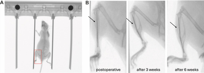
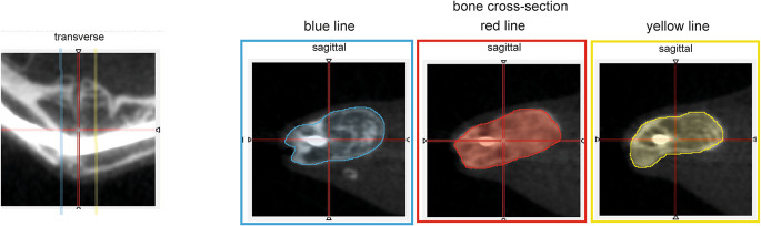
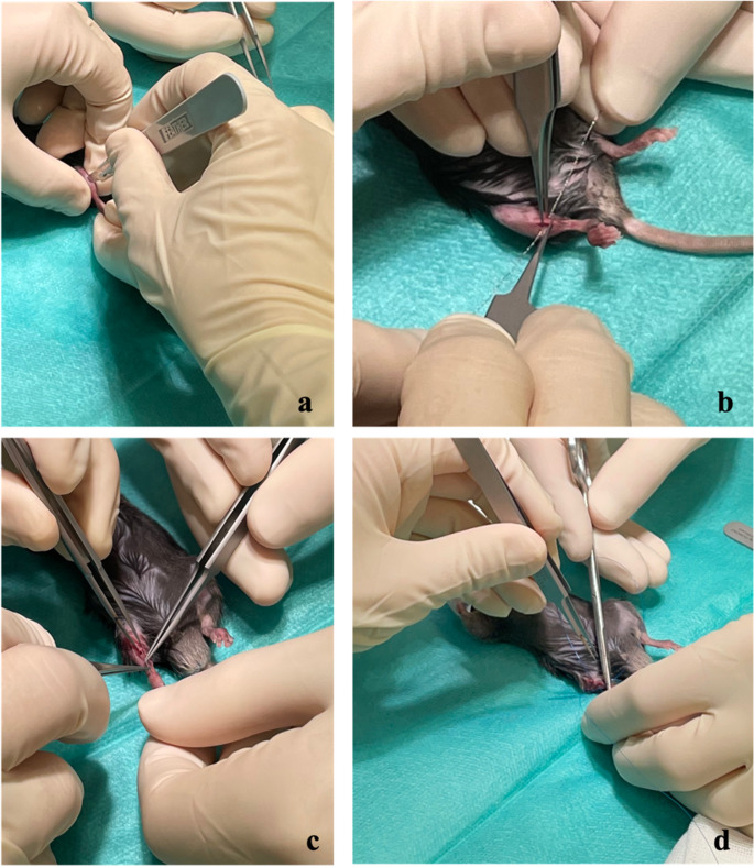
Methods: In 12 eight week-old wild type mice, an open tibial shaft fracture was simulated and stabilized with a retrograde over the fracture inserted intramedullary pin. X-rays confirmed the correct fracture localization and stabilization. After eight weeks of follow-up, the mice were euthanized. Fracture healing and biomechanical stability were analyzed in a micro-CT scan and in torsional load-to-failure tests.
Results: The whole operations lasted in mean eight min and 50 s. All mice recovered very quickly after the operative intervention and started using the operated leg again on the first postoperative day onwards if not earlier. No infections or failure of the stabilization occurred. All fractures healed completely within 8 weeks and substantial callus formation was confirmed in the micro-CT analysis. Biomechanically, higher torsional moment and stiffness were found for the operated tibia compared to the non-operated tibia in the same mouse.
Conclusion: The presented tibial fracture model with open osteotomy and retrograde pin insertion revealed minimal operative intervention and anesthesia, quick recovery and fracture healing with big callus formation. It is an easy to address fracture model for musculoskeletal research.
期刊介绍:
International Orthopaedics, the Official Journal of the Société Internationale de Chirurgie Orthopédique et de Traumatologie (SICOT) , publishes original papers from all over the world. The articles deal with clinical orthopaedic surgery or basic research directly connected with orthopaedic surgery. International Orthopaedics will also link all the members of SICOT by means of an insert that will be concerned with SICOT matters.
Finally, it is expected that news and information regarding all aspects of orthopaedic surgery, including meetings, panels, instructional courses, etc. will be brought to the attention of the readers.
Manuscripts submitted for publication must contain a statement to the effect that all human studies have been approved by the appropriate ethics committee and have therefore been performed in accordance with the ethical standards laid down in the 1964 Declaration of Helsinki. It should also be stated clearly in the text that all persons gave their informed consent prior to their inclusion in the study. Details that might disclose the identity of the subjects under study should be omitted.
Reports of animal experiments must state that the "Principles of laboratory animal care" (NIH publication No. 85-23, revised 1985) were followed, as well as specific national laws (e.g. the current version of the German Law on the Protection of Animals) where applicable.
The editors reserve the right to reject manuscripts that do not comply with the above-mentioned requirements. The author will be held responsible for false statements or for failure to fulfil the above-mentioned requirements.
 求助内容:
求助内容: 应助结果提醒方式:


