Kia Bayat, Aryan Seraj, Parisa Pooyan, Sepehr Feizi, Mozhgan Rezaei Kanavi, Marco A Zarbin, Hamid Ahmadieh
{"title":"Retinal and choroidal changes following corneal collagen cross-linking in keratoconus: a systematic review and meta-analysis of OCT and OCTA studies.","authors":"Kia Bayat, Aryan Seraj, Parisa Pooyan, Sepehr Feizi, Mozhgan Rezaei Kanavi, Marco A Zarbin, Hamid Ahmadieh","doi":"10.1186/s40942-025-00726-w","DOIUrl":null,"url":null,"abstract":"<p><p>Corneal collagen cross-linking (CXL) is widely used to halt the progression of keratoconus by biomechanically strengthening the corneal stroma; however, its potential effects on retina and choroid remain unclear. This systematic review and meta-analysis synthesized current evidence on structural and microvascular changes in the posterior segment following CXL in patients with keratoconus, assessed by optical coherence tomography (OCT) and OCT angiography (OCTA). A comprehensive search of PubMed, EMBASE, and Web of Science was performed up to May 24, 2025. Random-effects meta-analysis using Hedges' g was applied to pool quantitative data. In addition, studies that met the eligibility criteria but lacked sufficient data for quantitative synthesis were qualitatively assessed and included in the descriptive analysis. Ten studies involving 233 eyes from 215 keratoconus patients were included. Meta-analysis demonstrated no significant change in central macular thickness at 1 month (Hedges's g = -0.15; 95% confidence interval [CI]: -0.44 to 0.13; p = 0.30) or 6 months (Hedges's g = -0.12; 95% CI: -0.47 to 0.22; p = 0.48). Subfoveal choroidal thickness also remained unchanged at 1 month (Hedges's g = -0.14; 95% CI: -0.45 to 0.17; p = 0.37). Sensitivity analyses confirmed the robustness of these results. In the qualitative synthesis, parameters demonstrated overall stability, aside from a few exceptions. In conclusion, current evidence suggests that CXL does not result in clinically meaningful changes in posterior segment structure or microvasculature in keratoconus patients. These findings support the posterior segment safety of CXL.</p>","PeriodicalId":14289,"journal":{"name":"International Journal of Retina and Vitreous","volume":"11 1","pages":"97"},"PeriodicalIF":2.4000,"publicationDate":"2025-08-26","publicationTypes":"Journal Article","fieldsOfStudy":null,"isOpenAccess":false,"openAccessPdf":"https://www.ncbi.nlm.nih.gov/pmc/articles/PMC12379409/pdf/","citationCount":"0","resultStr":null,"platform":"Semanticscholar","paperid":null,"PeriodicalName":"International Journal of Retina and Vitreous","FirstCategoryId":"1085","ListUrlMain":"https://doi.org/10.1186/s40942-025-00726-w","RegionNum":0,"RegionCategory":null,"ArticlePicture":[],"TitleCN":null,"AbstractTextCN":null,"PMCID":null,"EPubDate":"","PubModel":"","JCR":"Q2","JCRName":"OPHTHALMOLOGY","Score":null,"Total":0}
引用次数: 0
Abstract
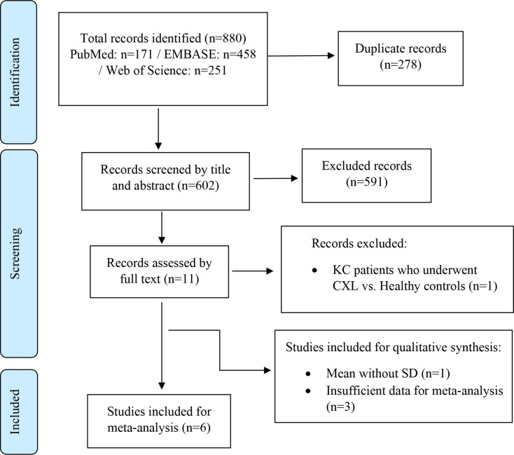
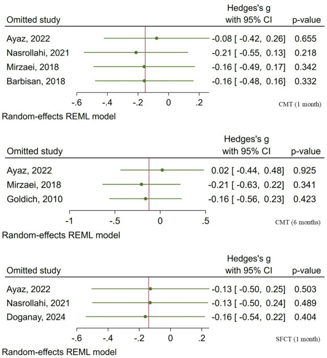
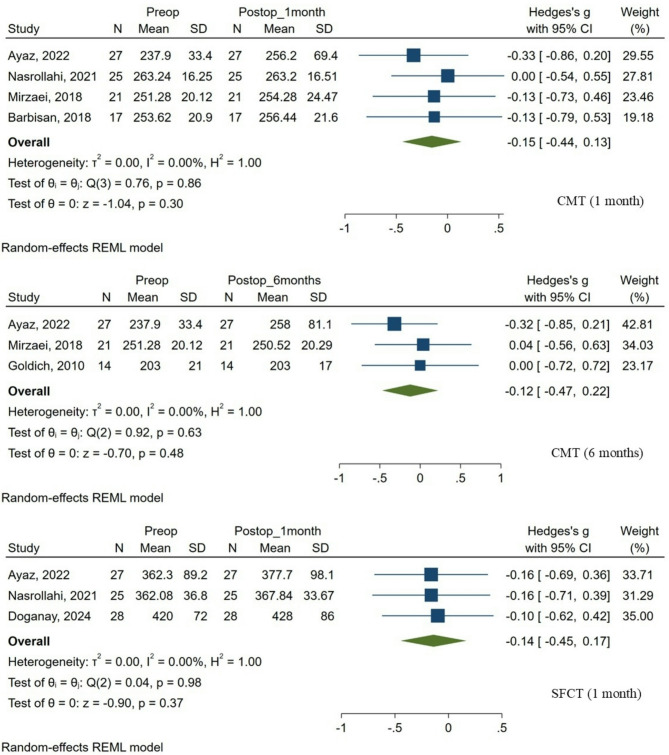
Corneal collagen cross-linking (CXL) is widely used to halt the progression of keratoconus by biomechanically strengthening the corneal stroma; however, its potential effects on retina and choroid remain unclear. This systematic review and meta-analysis synthesized current evidence on structural and microvascular changes in the posterior segment following CXL in patients with keratoconus, assessed by optical coherence tomography (OCT) and OCT angiography (OCTA). A comprehensive search of PubMed, EMBASE, and Web of Science was performed up to May 24, 2025. Random-effects meta-analysis using Hedges' g was applied to pool quantitative data. In addition, studies that met the eligibility criteria but lacked sufficient data for quantitative synthesis were qualitatively assessed and included in the descriptive analysis. Ten studies involving 233 eyes from 215 keratoconus patients were included. Meta-analysis demonstrated no significant change in central macular thickness at 1 month (Hedges's g = -0.15; 95% confidence interval [CI]: -0.44 to 0.13; p = 0.30) or 6 months (Hedges's g = -0.12; 95% CI: -0.47 to 0.22; p = 0.48). Subfoveal choroidal thickness also remained unchanged at 1 month (Hedges's g = -0.14; 95% CI: -0.45 to 0.17; p = 0.37). Sensitivity analyses confirmed the robustness of these results. In the qualitative synthesis, parameters demonstrated overall stability, aside from a few exceptions. In conclusion, current evidence suggests that CXL does not result in clinically meaningful changes in posterior segment structure or microvasculature in keratoconus patients. These findings support the posterior segment safety of CXL.
期刊介绍:
International Journal of Retina and Vitreous focuses on the ophthalmic subspecialty of vitreoretinal disorders. The journal presents original articles on new approaches to diagnosis, outcomes of clinical trials, innovations in pharmacological therapy and surgical techniques, as well as basic science advances that impact clinical practice. Topical areas include, but are not limited to: -Imaging of the retina, choroid and vitreous -Innovations in optical coherence tomography (OCT) -Small-gauge vitrectomy, retinal detachment, chromovitrectomy -Electroretinography (ERG), microperimetry, other functional tests -Intraocular tumors -Retinal pharmacotherapy & drug delivery -Diabetic retinopathy & other vascular diseases -Age-related macular degeneration (AMD) & other macular entities

 求助内容:
求助内容: 应助结果提醒方式:


