Tisha Thomas Menacherry, V Rajesh Prabu, Jagadeesh Kumar Reddy, Suruthi Nagarajan, Mithun Thulasidas
{"title":"Comparison of retinal microvasculature in anisometropic amblyopic and healthy eyes using optical coherence tomography angiography.","authors":"Tisha Thomas Menacherry, V Rajesh Prabu, Jagadeesh Kumar Reddy, Suruthi Nagarajan, Mithun Thulasidas","doi":"10.4103/IJO.IJO_1047_25","DOIUrl":null,"url":null,"abstract":"<p><strong>Purpose: </strong>To compare the optical coherence tomography angiography (OCTA) findings of retinal microvasculature in anisometropic amblyopic eyes with fellow eyes.</p><p><strong>Methods: </strong>A comparative observational study was conducted in children with unilateral anisometropic amblyopia to compare the OCTA findings of retinal microvasculature between amblyopic and normal fellow eyes. Major outcome measures included superficial vessel density, deep vessel density, and foveal avascular zone (FAZ) parameters (area, perimetry, acircularity index) of amblyopic eye and fellow eye on OCTA.</p><p><strong>Results: </strong>The study involved 40 children with a mean age of 10.25 ± 3.07 years. Amblyopic eyes showed significantly reduced vessel density in the superficial and deep plexuses compared to fellow eyes (P < 0.05). Reductions were more pronounced in severe amblyopia, with significant foveal differences in superficial (P = 0.04) and deep layers (P = 0.02). A significant negative correlation was observed between best corrected visual acuity and vessel density.</p><p><strong>Conclusion: </strong>This study found reduced retinal vessel density in anisometropic amblyopic eyes, particularly in the superficial and deep capillary plexuses. Vessel density correlates with the severity of amblyopia, suggesting its potential as a biomarker for disease progression and severity.</p>","PeriodicalId":13329,"journal":{"name":"Indian Journal of Ophthalmology","volume":"73 9","pages":"1376-1382"},"PeriodicalIF":1.8000,"publicationDate":"2025-09-01","publicationTypes":"Journal Article","fieldsOfStudy":null,"isOpenAccess":false,"openAccessPdf":"https://www.ncbi.nlm.nih.gov/pmc/articles/PMC12448494/pdf/","citationCount":"0","resultStr":null,"platform":"Semanticscholar","paperid":null,"PeriodicalName":"Indian Journal of Ophthalmology","FirstCategoryId":"3","ListUrlMain":"https://doi.org/10.4103/IJO.IJO_1047_25","RegionNum":4,"RegionCategory":"医学","ArticlePicture":[],"TitleCN":null,"AbstractTextCN":null,"PMCID":null,"EPubDate":"2025/8/29 0:00:00","PubModel":"Epub","JCR":"Q2","JCRName":"OPHTHALMOLOGY","Score":null,"Total":0}
引用次数: 0
Abstract
Purpose: To compare the optical coherence tomography angiography (OCTA) findings of retinal microvasculature in anisometropic amblyopic eyes with fellow eyes.
Methods: A comparative observational study was conducted in children with unilateral anisometropic amblyopia to compare the OCTA findings of retinal microvasculature between amblyopic and normal fellow eyes. Major outcome measures included superficial vessel density, deep vessel density, and foveal avascular zone (FAZ) parameters (area, perimetry, acircularity index) of amblyopic eye and fellow eye on OCTA.
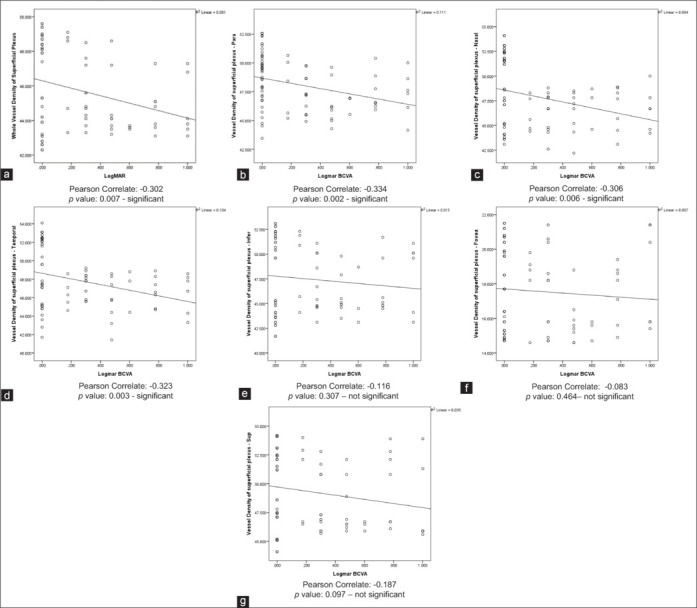
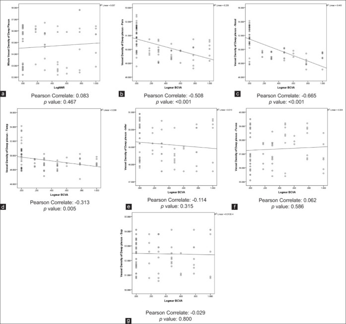
Results: The study involved 40 children with a mean age of 10.25 ± 3.07 years. Amblyopic eyes showed significantly reduced vessel density in the superficial and deep plexuses compared to fellow eyes (P < 0.05). Reductions were more pronounced in severe amblyopia, with significant foveal differences in superficial (P = 0.04) and deep layers (P = 0.02). A significant negative correlation was observed between best corrected visual acuity and vessel density.
Conclusion: This study found reduced retinal vessel density in anisometropic amblyopic eyes, particularly in the superficial and deep capillary plexuses. Vessel density correlates with the severity of amblyopia, suggesting its potential as a biomarker for disease progression and severity.
期刊介绍:
Indian Journal of Ophthalmology covers clinical, experimental, basic science research and translational research studies related to medical, ethical and social issues in field of ophthalmology and vision science. Articles with clinical interest and implications will be given preference.
 求助内容:
求助内容: 应助结果提醒方式:


