{"title":"Multi-spin Redox Sensor for Electron Paramagnetic Resonance Studies of Tissue Redox State <i>In Vivo</i>: Validation of Data With a Conventional Spin Probe.","authors":"Dessislava Lazarova, Biliana Nikolova, Zhivko Zhelev, Plamen Getsov, Milka Mileva, Rumiana Bakalova, Ichio Aoki","doi":"10.21873/invivo.14069","DOIUrl":null,"url":null,"abstract":"<p><strong>Background/aim: </strong>Redox imaging is one of the fastest growing areas in diagnostics of pathologies accompanied by redox imbalance. We describe a multi-spin redox sensor (RS) and its application for redox imaging in mice using electron paramagnetic resonance (EPR) spectroscopy.</p><p><strong>Materials and methods: </strong>The probe is composed of a quantum dot functionalized with a cyclodextrin shell, conjugated with nitroxide residues (TEMPO) and triphenylphosphonium to achieve intracellular delivery. The data were validated with a conventional spin probe, mito-TEMPO. Nitroxide probe (RS or mito-TEMPO) was injected intravenously in mice under anesthesia. Blood samples were collected from the tail vein at four time intervals within 2 hours and subjected immediately to analysis using EPR spectroscopy. Two hours after injection, mice were sacrificed, five organs were isolated, and tissue homogenates were prepared and subjected to EPR analysis.</p><p><strong>Results: </strong>RS had the same EPR contrast and significantly higher MRI contrast compared to mito-TEMPO. RS circulated longer in the bloodstream than mito-TEMPO, with both substances undergoing reduction in the blood. The distribution of RS and mito-TEMPO in different organs was equal, except for the brain. No adverse side effects were found in mice at the selected dose.</p><p><strong>Conclusion: </strong>Using a non-toxic nanoparticle in the chemical structure of RS as a carrier of nitroxide residues would enhance its translational relevance.</p>","PeriodicalId":13364,"journal":{"name":"In vivo","volume":"39 5","pages":"2703-2710"},"PeriodicalIF":1.8000,"publicationDate":"2025-09-01","publicationTypes":"Journal Article","fieldsOfStudy":null,"isOpenAccess":false,"openAccessPdf":"https://www.ncbi.nlm.nih.gov/pmc/articles/PMC12396052/pdf/","citationCount":"0","resultStr":null,"platform":"Semanticscholar","paperid":null,"PeriodicalName":"In vivo","FirstCategoryId":"3","ListUrlMain":"https://doi.org/10.21873/invivo.14069","RegionNum":4,"RegionCategory":"医学","ArticlePicture":[],"TitleCN":null,"AbstractTextCN":null,"PMCID":null,"EPubDate":"","PubModel":"","JCR":"Q3","JCRName":"MEDICINE, RESEARCH & EXPERIMENTAL","Score":null,"Total":0}
引用次数: 0
Abstract
Background/aim: Redox imaging is one of the fastest growing areas in diagnostics of pathologies accompanied by redox imbalance. We describe a multi-spin redox sensor (RS) and its application for redox imaging in mice using electron paramagnetic resonance (EPR) spectroscopy.
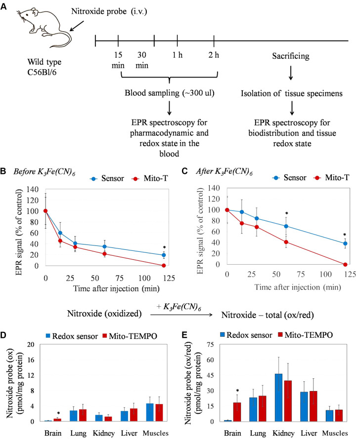
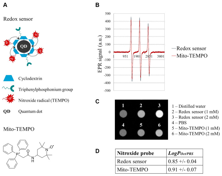
Materials and methods: The probe is composed of a quantum dot functionalized with a cyclodextrin shell, conjugated with nitroxide residues (TEMPO) and triphenylphosphonium to achieve intracellular delivery. The data were validated with a conventional spin probe, mito-TEMPO. Nitroxide probe (RS or mito-TEMPO) was injected intravenously in mice under anesthesia. Blood samples were collected from the tail vein at four time intervals within 2 hours and subjected immediately to analysis using EPR spectroscopy. Two hours after injection, mice were sacrificed, five organs were isolated, and tissue homogenates were prepared and subjected to EPR analysis.
Results: RS had the same EPR contrast and significantly higher MRI contrast compared to mito-TEMPO. RS circulated longer in the bloodstream than mito-TEMPO, with both substances undergoing reduction in the blood. The distribution of RS and mito-TEMPO in different organs was equal, except for the brain. No adverse side effects were found in mice at the selected dose.
Conclusion: Using a non-toxic nanoparticle in the chemical structure of RS as a carrier of nitroxide residues would enhance its translational relevance.
期刊介绍:
IN VIVO is an international peer-reviewed journal designed to bring together original high quality works and reviews on experimental and clinical biomedical research within the frames of physiology, pathology and disease management.
The topics of IN VIVO include: 1. Experimental development and application of new diagnostic and therapeutic procedures; 2. Pharmacological and toxicological evaluation of new drugs, drug combinations and drug delivery systems; 3. Clinical trials; 4. Development and characterization of models of biomedical research; 5. Cancer diagnosis and treatment; 6. Immunotherapy and vaccines; 7. Radiotherapy, Imaging; 8. Tissue engineering, Regenerative medicine; 9. Carcinogenesis.

 求助内容:
求助内容: 应助结果提醒方式:


