Yuki Shinohara, Jun Nishio, Shizuhide Nakayama, Mikiko Aoki
{"title":"Lipofibromatosis Revisited.","authors":"Yuki Shinohara, Jun Nishio, Shizuhide Nakayama, Mikiko Aoki","doi":"10.21873/invivo.14054","DOIUrl":null,"url":null,"abstract":"<p><p>Lipofibromatosis (LPF) is a locally aggressive but non-metastasizing mesenchymal tumor that primarily occurs in the hands and feet of infants and young children. It typically presents as a slow-growing, painless, poorly demarcated subcutaneous mass. Magnetic resonance imaging reveals the lesion to be a poorly defined mass with a mixture of adipose and fibrous components. Variable enhancement is seen after intravenous contrast administration. Histologically, LPF displays a distinctive admixture of mature adipose tissue and short fascicles of bland spindle cells. By immunohistochemistry, the spindle cells are moderately or diffusely positive for CD34 and CD99, focally positive for smooth muscle actin but typically negative for S-100 protein, desmin, β-catenin and pan-tropomyosin receptor kinase (TRK). Recent molecular studies have shown a variety of fusions involving epidermal growth factor receptor (EGFR) ligands or EGFR itself or other receptor tyrosine kinases, suggesting a shared deregulation of the phosphatidylinositol 3-kinase (PI3K)/AKT/mammalian target of the rapamycin (mTOR) pathway. Complete surgical excision with preservation of adjacent neurovascular structures is the treatment of choice for LPF. This review provides an updated overview of the clinical, radiological, histological, immunohistochemical, cytogenetic and molecular genetic features of LPF and discusses the relationship to LPF-like neural tumor.</p>","PeriodicalId":13364,"journal":{"name":"In vivo","volume":"39 5","pages":"2512-2516"},"PeriodicalIF":1.8000,"publicationDate":"2025-09-01","publicationTypes":"Journal Article","fieldsOfStudy":null,"isOpenAccess":false,"openAccessPdf":"https://www.ncbi.nlm.nih.gov/pmc/articles/PMC12396084/pdf/","citationCount":"0","resultStr":null,"platform":"Semanticscholar","paperid":null,"PeriodicalName":"In vivo","FirstCategoryId":"3","ListUrlMain":"https://doi.org/10.21873/invivo.14054","RegionNum":4,"RegionCategory":"医学","ArticlePicture":[],"TitleCN":null,"AbstractTextCN":null,"PMCID":null,"EPubDate":"","PubModel":"","JCR":"Q3","JCRName":"MEDICINE, RESEARCH & EXPERIMENTAL","Score":null,"Total":0}
引用次数: 0
Abstract
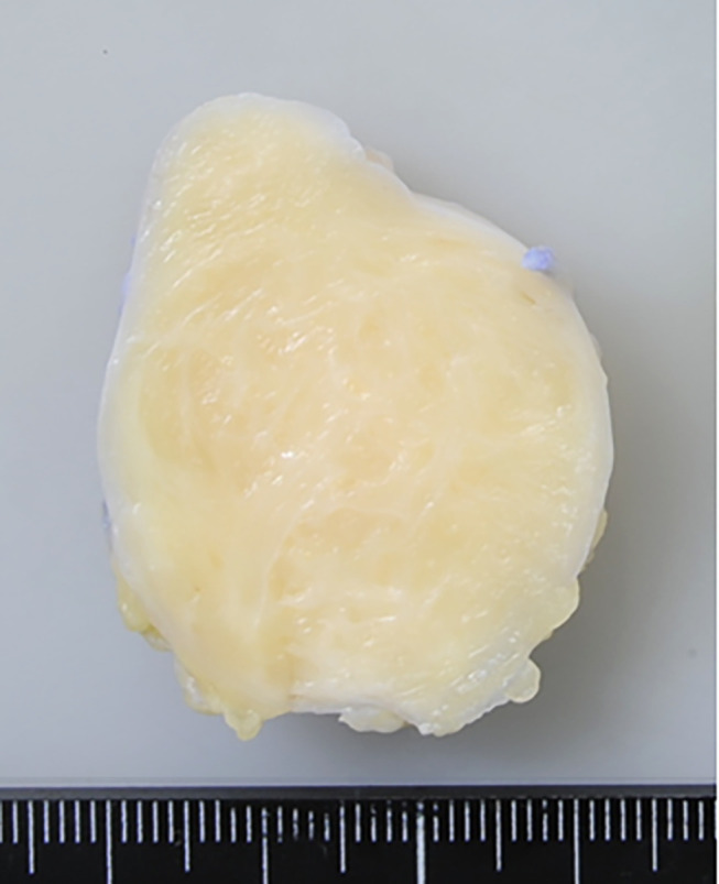
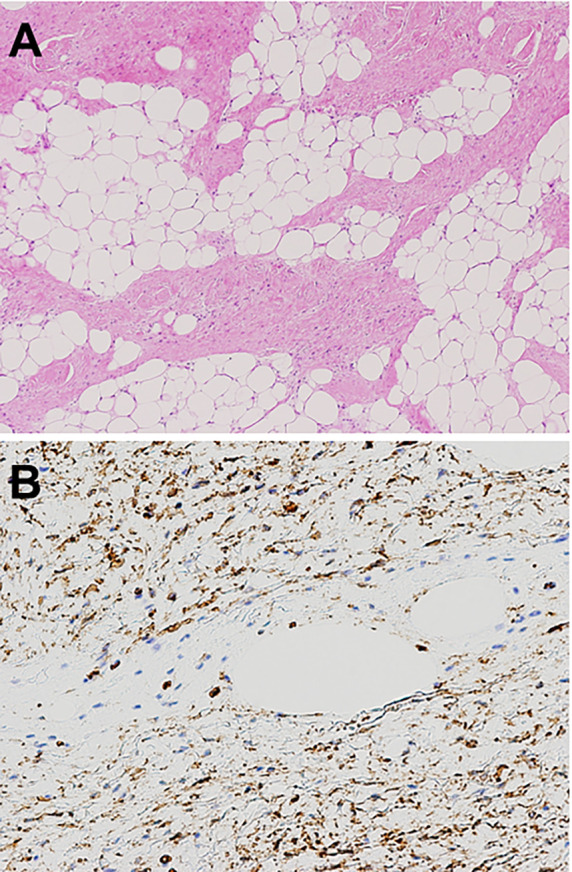
Lipofibromatosis (LPF) is a locally aggressive but non-metastasizing mesenchymal tumor that primarily occurs in the hands and feet of infants and young children. It typically presents as a slow-growing, painless, poorly demarcated subcutaneous mass. Magnetic resonance imaging reveals the lesion to be a poorly defined mass with a mixture of adipose and fibrous components. Variable enhancement is seen after intravenous contrast administration. Histologically, LPF displays a distinctive admixture of mature adipose tissue and short fascicles of bland spindle cells. By immunohistochemistry, the spindle cells are moderately or diffusely positive for CD34 and CD99, focally positive for smooth muscle actin but typically negative for S-100 protein, desmin, β-catenin and pan-tropomyosin receptor kinase (TRK). Recent molecular studies have shown a variety of fusions involving epidermal growth factor receptor (EGFR) ligands or EGFR itself or other receptor tyrosine kinases, suggesting a shared deregulation of the phosphatidylinositol 3-kinase (PI3K)/AKT/mammalian target of the rapamycin (mTOR) pathway. Complete surgical excision with preservation of adjacent neurovascular structures is the treatment of choice for LPF. This review provides an updated overview of the clinical, radiological, histological, immunohistochemical, cytogenetic and molecular genetic features of LPF and discusses the relationship to LPF-like neural tumor.
期刊介绍:
IN VIVO is an international peer-reviewed journal designed to bring together original high quality works and reviews on experimental and clinical biomedical research within the frames of physiology, pathology and disease management.
The topics of IN VIVO include: 1. Experimental development and application of new diagnostic and therapeutic procedures; 2. Pharmacological and toxicological evaluation of new drugs, drug combinations and drug delivery systems; 3. Clinical trials; 4. Development and characterization of models of biomedical research; 5. Cancer diagnosis and treatment; 6. Immunotherapy and vaccines; 7. Radiotherapy, Imaging; 8. Tissue engineering, Regenerative medicine; 9. Carcinogenesis.

 求助内容:
求助内容: 应助结果提醒方式:


