{"title":"Development and Characterization of a Murine Lung Adenocarcinoma Cell Line With High Thoracic Pleural Metastatic Potential.","authors":"Liwei Liao, Weidong Xu, Jia Li, Jiaye Li, Rui Li, Ruixia Li, Chang Li, Ziwen Zheng, Mingming Deng, Jinrui Miao, Zilin Wang, Qin Zhang, Yiding Bian, Kai Wang, Han Wang, Gang Hou","doi":"10.21873/invivo.14067","DOIUrl":null,"url":null,"abstract":"<p><strong>Background/aim: </strong>Pleural metastasis and malignant pleural effusion (MPE) are common complications of lung adenocarcinoma. Patients with MPE have poor outcomes, with overall survival ranging from 5 to 11.4 months. The lack of established cell lines and stable animal models of pleural metastasis has limited studies on the underlying mechanisms of MPE development. In this study, we aimed to develop a murine lung adenocarcinoma cell line with high thoracic pleural metastatic potential.</p><p><strong>Materials and methods: </strong>Luciferase-tagged Lewis lung carcinoma (LLC) cells were implanted into the pleural cavity of C57bl/6 mice, with five rounds of subsequent extraction from pleural foci and reinjection into the pleural cavity. The metastatic properties of the established cell line were verified <i>in vivo</i> by evaluating the metastatic burden and MPE volume (n=5). <i>In vitro</i>, the metastatic ability of cell lines was assessed by scratch assay, transwell migration assay, cell-matrix adhesion assay and cell-cell adhesion assay (3-5 replicates). The transcription profile was characterized by mRNA sequencing. Differential analysis and KEGG enrichment were performed to show their distinctions. Differential genes were verified by quantitative real-time PCR (qPCR).</p><p><strong>Results: </strong>An LLC subpopulation with high thoracic pleural metastatic potential was generated and named LLC-PLM. <i>In vivo</i>, compared with parental LLC (LLC-P), LLC-PLM demonstrated a greater incidence of MPE and greater MPE volumes. <i>In vitro</i>, LLC-PLM demonstrated increased metastatic capacity and augmented adhesion capacities, compared to LLC-P. Transcriptomic analysis revealed that pathways related to adhesion, migration, and membrane signaling were notably enriched and activated in LLC-PLM cells. Relative genes were obviously activated, including Lamc2, Col4a3, Col6a3, Col1a1, Itga2 and Itga1.</p><p><strong>Conclusion: </strong>We successfully established a murine cell line LLC-PLM that can serve as a valuable tool for studying pleural metastasis and MPE.</p>","PeriodicalId":13364,"journal":{"name":"In vivo","volume":"39 5","pages":"2669-2680"},"PeriodicalIF":1.8000,"publicationDate":"2025-09-01","publicationTypes":"Journal Article","fieldsOfStudy":null,"isOpenAccess":false,"openAccessPdf":"https://www.ncbi.nlm.nih.gov/pmc/articles/PMC12396039/pdf/","citationCount":"0","resultStr":null,"platform":"Semanticscholar","paperid":null,"PeriodicalName":"In vivo","FirstCategoryId":"3","ListUrlMain":"https://doi.org/10.21873/invivo.14067","RegionNum":4,"RegionCategory":"医学","ArticlePicture":[],"TitleCN":null,"AbstractTextCN":null,"PMCID":null,"EPubDate":"","PubModel":"","JCR":"Q3","JCRName":"MEDICINE, RESEARCH & EXPERIMENTAL","Score":null,"Total":0}
引用次数: 0
Abstract
Background/aim: Pleural metastasis and malignant pleural effusion (MPE) are common complications of lung adenocarcinoma. Patients with MPE have poor outcomes, with overall survival ranging from 5 to 11.4 months. The lack of established cell lines and stable animal models of pleural metastasis has limited studies on the underlying mechanisms of MPE development. In this study, we aimed to develop a murine lung adenocarcinoma cell line with high thoracic pleural metastatic potential.
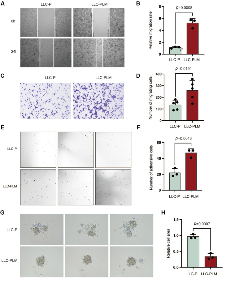
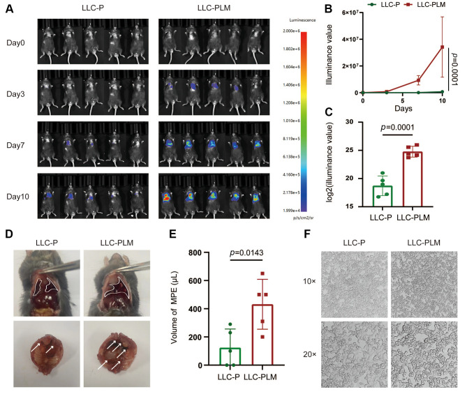
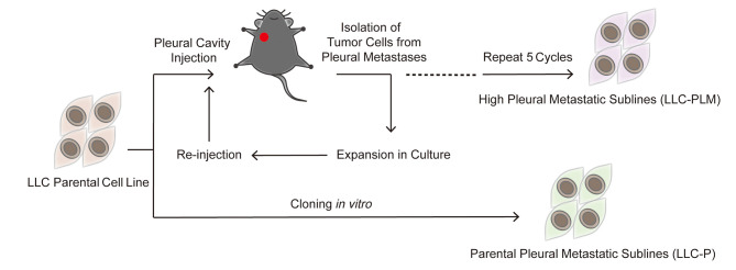
Materials and methods: Luciferase-tagged Lewis lung carcinoma (LLC) cells were implanted into the pleural cavity of C57bl/6 mice, with five rounds of subsequent extraction from pleural foci and reinjection into the pleural cavity. The metastatic properties of the established cell line were verified in vivo by evaluating the metastatic burden and MPE volume (n=5). In vitro, the metastatic ability of cell lines was assessed by scratch assay, transwell migration assay, cell-matrix adhesion assay and cell-cell adhesion assay (3-5 replicates). The transcription profile was characterized by mRNA sequencing. Differential analysis and KEGG enrichment were performed to show their distinctions. Differential genes were verified by quantitative real-time PCR (qPCR).
Results: An LLC subpopulation with high thoracic pleural metastatic potential was generated and named LLC-PLM. In vivo, compared with parental LLC (LLC-P), LLC-PLM demonstrated a greater incidence of MPE and greater MPE volumes. In vitro, LLC-PLM demonstrated increased metastatic capacity and augmented adhesion capacities, compared to LLC-P. Transcriptomic analysis revealed that pathways related to adhesion, migration, and membrane signaling were notably enriched and activated in LLC-PLM cells. Relative genes were obviously activated, including Lamc2, Col4a3, Col6a3, Col1a1, Itga2 and Itga1.
Conclusion: We successfully established a murine cell line LLC-PLM that can serve as a valuable tool for studying pleural metastasis and MPE.
期刊介绍:
IN VIVO is an international peer-reviewed journal designed to bring together original high quality works and reviews on experimental and clinical biomedical research within the frames of physiology, pathology and disease management.
The topics of IN VIVO include: 1. Experimental development and application of new diagnostic and therapeutic procedures; 2. Pharmacological and toxicological evaluation of new drugs, drug combinations and drug delivery systems; 3. Clinical trials; 4. Development and characterization of models of biomedical research; 5. Cancer diagnosis and treatment; 6. Immunotherapy and vaccines; 7. Radiotherapy, Imaging; 8. Tissue engineering, Regenerative medicine; 9. Carcinogenesis.

 求助内容:
求助内容: 应助结果提醒方式:


