Mohammad Amin Shayestehpour, Martin G Gregersen, Ola Saatvedt, Øystein Bjelland, Marius Molund
{"title":"A Pilot Validation Study of a Biomechanical Simulation Model for Rotational Ankle Injuries Using Robotic Cadaveric Testing.","authors":"Mohammad Amin Shayestehpour, Martin G Gregersen, Ola Saatvedt, Øystein Bjelland, Marius Molund","doi":"10.1177/24730114251356497","DOIUrl":null,"url":null,"abstract":"<p><strong>Background: </strong>Deltoid ligament injuries occur in specific sequences during rotational ankle trauma, yet the current understanding of these sequences may be flawed. Computer modeling offers a new method for assessing ligament behavior under rotational injury mechanisms.</p><p><strong>Methods: </strong>A biomechanical computer simulation model was developed using AnyBody Modeling Software to evaluate ligament strain in rotational ankle injuries. Experimental data from a cadaveric study involving 15 human ankle specimens subjected to various loading conditions were used to identify the model parameters. After parameter identification from uninjured cadaveric data, we simulated Supination-External Rotation (SER) stage 2-4b injury model by removing the corresponding ligaments. Validation was performed by comparing the model predictions against the biomechanical experimental data.</p><p><strong>Results: </strong>The computer model replicated experimental findings, with correlation coefficients ranging from 0.81 to 0.99 across all injury stages and loading conditions. Furthermore, tension in the deep posterior tibiotalar ligament (DPTTL) progressively increased from SER2 to SER4a but remained unchanged in the SER2 phase. The model effectively captured progressive ligament strain and changes in medial clear space during injury progression.</p><p><strong>Conclusion: </strong>This study presents and validates an early-stage biomechanical simulation model for rotational ankle injuries, providing a novel tool for examining ligament biomechanics and injury mechanisms.</p><p><strong>Clinical relevance: </strong>Our model offers insights that were previously unattainable through cadaveric or clinical studies by simulating ligament strain during injuries. This can assist in generating hypotheses, enhance injury detection, refine treatment strategies, and may challenge existing classification systems.</p>","PeriodicalId":12429,"journal":{"name":"Foot & Ankle Orthopaedics","volume":"10 3","pages":"24730114251356497"},"PeriodicalIF":0.0000,"publicationDate":"2025-08-20","publicationTypes":"Journal Article","fieldsOfStudy":null,"isOpenAccess":false,"openAccessPdf":"https://www.ncbi.nlm.nih.gov/pmc/articles/PMC12368408/pdf/","citationCount":"0","resultStr":null,"platform":"Semanticscholar","paperid":null,"PeriodicalName":"Foot & Ankle Orthopaedics","FirstCategoryId":"1085","ListUrlMain":"https://doi.org/10.1177/24730114251356497","RegionNum":0,"RegionCategory":null,"ArticlePicture":[],"TitleCN":null,"AbstractTextCN":null,"PMCID":null,"EPubDate":"2025/7/1 0:00:00","PubModel":"eCollection","JCR":"","JCRName":"","Score":null,"Total":0}
引用次数: 0
Abstract
Background: Deltoid ligament injuries occur in specific sequences during rotational ankle trauma, yet the current understanding of these sequences may be flawed. Computer modeling offers a new method for assessing ligament behavior under rotational injury mechanisms.
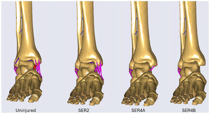
Methods: A biomechanical computer simulation model was developed using AnyBody Modeling Software to evaluate ligament strain in rotational ankle injuries. Experimental data from a cadaveric study involving 15 human ankle specimens subjected to various loading conditions were used to identify the model parameters. After parameter identification from uninjured cadaveric data, we simulated Supination-External Rotation (SER) stage 2-4b injury model by removing the corresponding ligaments. Validation was performed by comparing the model predictions against the biomechanical experimental data.
Results: The computer model replicated experimental findings, with correlation coefficients ranging from 0.81 to 0.99 across all injury stages and loading conditions. Furthermore, tension in the deep posterior tibiotalar ligament (DPTTL) progressively increased from SER2 to SER4a but remained unchanged in the SER2 phase. The model effectively captured progressive ligament strain and changes in medial clear space during injury progression.
Conclusion: This study presents and validates an early-stage biomechanical simulation model for rotational ankle injuries, providing a novel tool for examining ligament biomechanics and injury mechanisms.
Clinical relevance: Our model offers insights that were previously unattainable through cadaveric or clinical studies by simulating ligament strain during injuries. This can assist in generating hypotheses, enhance injury detection, refine treatment strategies, and may challenge existing classification systems.

 求助内容:
求助内容: 应助结果提醒方式:


