Giuseppe D'Albis, Marta Forte, Laura Stef, Diana Ramona Feier, Victor Diaz-Flores García, Massimo Corsalini, Saverio Capodiferro
{"title":"A Digital Workflow for Virtual Articulator Mounting Using Face Scan and Facebow Capture: A Proof-of-Concept.","authors":"Giuseppe D'Albis, Marta Forte, Laura Stef, Diana Ramona Feier, Victor Diaz-Flores García, Massimo Corsalini, Saverio Capodiferro","doi":"10.3390/dj13080378","DOIUrl":null,"url":null,"abstract":"<p><p><b>Objectives:</b> This article introduces a digital technique for virtual articulator mounting by employing the scan of a facebow worn by the patient as a virtual reference. <b>Methods:</b> The digital technique enables the transfer of the maxillary arch orientation relative to the cranial base into a CAD-CAM environment (Ceramill Mind; AmannGirrbach), without the need for ionizing radiation or identification of facial landmarks. By digitally aligning the intraoral scans of the dental arches (Trios 4; 3Shape) with a 3D facial scan and the scanned facebow in position (Artex; AmannGirrbach), clinicians can reproduce the cranium-to-maxilla spatial relationship accurately and intuitively. <b>Results:</b> This radiation-free protocol provides virtual cross-mounting and allows for the use of a semi-adjustable articulator within common CAD-CAM software. <b>Conclusions:</b> Given that intraoral scanners, facial scanners, and design software with articulator simulation are becoming more available in modern clinical workflows, this method introduced here could be a viable radiation-free and easy-to-use alternative. However, larger cohorts and standardized testing protocols are needed to determine its clinical reproducibility and reliability.</p>","PeriodicalId":11269,"journal":{"name":"Dentistry Journal","volume":"13 8","pages":""},"PeriodicalIF":3.1000,"publicationDate":"2025-08-20","publicationTypes":"Journal Article","fieldsOfStudy":null,"isOpenAccess":false,"openAccessPdf":"https://www.ncbi.nlm.nih.gov/pmc/articles/PMC12384779/pdf/","citationCount":"0","resultStr":null,"platform":"Semanticscholar","paperid":null,"PeriodicalName":"Dentistry Journal","FirstCategoryId":"1085","ListUrlMain":"https://doi.org/10.3390/dj13080378","RegionNum":0,"RegionCategory":null,"ArticlePicture":[],"TitleCN":null,"AbstractTextCN":null,"PMCID":null,"EPubDate":"","PubModel":"","JCR":"Q2","JCRName":"DENTISTRY, ORAL SURGERY & MEDICINE","Score":null,"Total":0}
引用次数: 0
Abstract
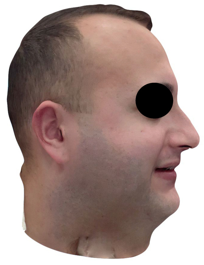
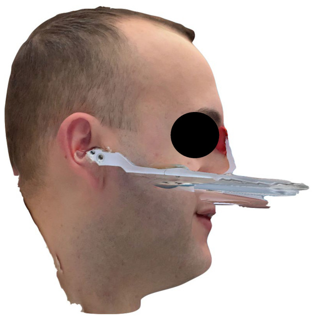
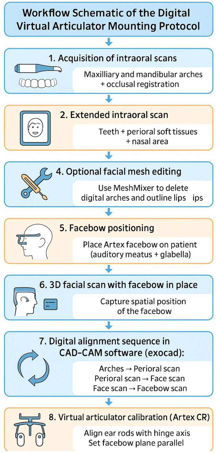
Objectives: This article introduces a digital technique for virtual articulator mounting by employing the scan of a facebow worn by the patient as a virtual reference. Methods: The digital technique enables the transfer of the maxillary arch orientation relative to the cranial base into a CAD-CAM environment (Ceramill Mind; AmannGirrbach), without the need for ionizing radiation or identification of facial landmarks. By digitally aligning the intraoral scans of the dental arches (Trios 4; 3Shape) with a 3D facial scan and the scanned facebow in position (Artex; AmannGirrbach), clinicians can reproduce the cranium-to-maxilla spatial relationship accurately and intuitively. Results: This radiation-free protocol provides virtual cross-mounting and allows for the use of a semi-adjustable articulator within common CAD-CAM software. Conclusions: Given that intraoral scanners, facial scanners, and design software with articulator simulation are becoming more available in modern clinical workflows, this method introduced here could be a viable radiation-free and easy-to-use alternative. However, larger cohorts and standardized testing protocols are needed to determine its clinical reproducibility and reliability.

 求助内容:
求助内容: 应助结果提醒方式:


