Comparison of the Marginal Integrity of Sectional Non-Invasive Laminate Veneers Versus Sectional Minimally Invasive Laminate Veneers Under Artificial Aging.
Polykarpos Papanagiotou, Phophi Kamposiora, George Papavasiliou, Spiros Zinelis
{"title":"Comparison of the Marginal Integrity of Sectional Non-Invasive Laminate Veneers Versus Sectional Minimally Invasive Laminate Veneers Under Artificial Aging.","authors":"Polykarpos Papanagiotou, Phophi Kamposiora, George Papavasiliou, Spiros Zinelis","doi":"10.3390/dj13080358","DOIUrl":null,"url":null,"abstract":"<p><p><b>Background/Objectives</b>: To compare the marginal integrity of sectional non-invasive laminate veneers versus sectional minimally invasive laminate veneers. <b>Methods</b>: A total of eighty (80) intact maxillary and mandibular frontal teeth (central incisors) were randomly divided into four groups (<i>n</i> = 20). Two groups received non-invasive veneers made of feldspathic porcelain (Feldspathic Non-Invasive-FNI) and lithium disilicate (Lithium Disilicate Non-Invasive-LNI) accordingly. Groups FP and LP received minimally invasive veneers manufactured by feldspathic porcelain and lithium disilicate, respectively. Following cementation, three grooves with mesio-distal orientation on the labial surface of teeth of each sample, at the incisal, middle, and cervical third, were made. Before and after artificial aging, the mesio-distal distance between the end of the groove on the tooth and the edge of each veneer was measured in micrometers (μm) employing an SEM immediately after cementation (T<sub>0</sub>), after simulated artificial aging equivalent to four months of everyday brushing (T<sub>1</sub>), and after twelve months of everyday brushing (2 times per day) (T<sub>2</sub>) to identify the wear of veneers. In the same manner, the horizontal dimension of the cement layer extending from its edge till the margin of the veneer was measured for all the groups at T<sub>0</sub>, T<sub>1</sub>, and T<sub>2</sub>, respectively. The statistical analysis was performed employing non-parametric Kruskal-Wallis ANOVA and Dunn's test. <b>Results</b>: No significant differences from T<sub>0</sub> to T<sub>1</sub> and from T<sub>1</sub> to T<sub>2</sub>, as well as from T<sub>0</sub> to T<sub>2</sub>, were identified for all the groups tested. No significant differences were allocated among all groups for the dimensional changes in the cement. <b>Conclusions</b>: All the groups responded similarly to aging factors, regardless of the non-invasive or minimally invasive approach, or the material used to fabricate the veneers.</p>","PeriodicalId":11269,"journal":{"name":"Dentistry Journal","volume":"13 8","pages":""},"PeriodicalIF":3.1000,"publicationDate":"2025-08-07","publicationTypes":"Journal Article","fieldsOfStudy":null,"isOpenAccess":false,"openAccessPdf":"https://www.ncbi.nlm.nih.gov/pmc/articles/PMC12384641/pdf/","citationCount":"0","resultStr":null,"platform":"Semanticscholar","paperid":null,"PeriodicalName":"Dentistry Journal","FirstCategoryId":"1085","ListUrlMain":"https://doi.org/10.3390/dj13080358","RegionNum":0,"RegionCategory":null,"ArticlePicture":[],"TitleCN":null,"AbstractTextCN":null,"PMCID":null,"EPubDate":"","PubModel":"","JCR":"Q2","JCRName":"DENTISTRY, ORAL SURGERY & MEDICINE","Score":null,"Total":0}
引用次数: 0
Abstract
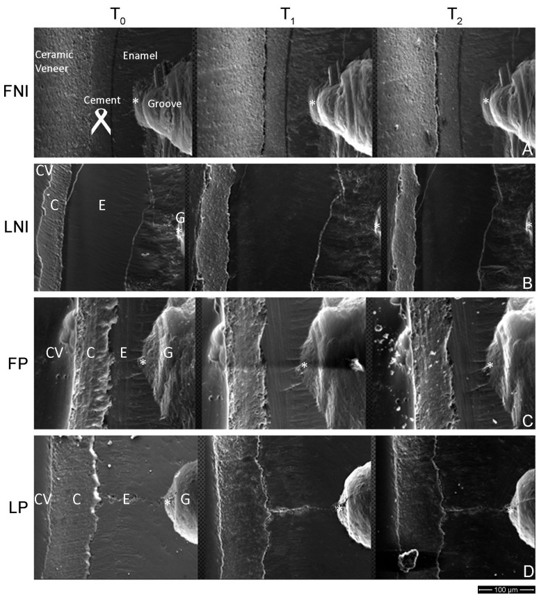
Background/Objectives: To compare the marginal integrity of sectional non-invasive laminate veneers versus sectional minimally invasive laminate veneers. Methods: A total of eighty (80) intact maxillary and mandibular frontal teeth (central incisors) were randomly divided into four groups (n = 20). Two groups received non-invasive veneers made of feldspathic porcelain (Feldspathic Non-Invasive-FNI) and lithium disilicate (Lithium Disilicate Non-Invasive-LNI) accordingly. Groups FP and LP received minimally invasive veneers manufactured by feldspathic porcelain and lithium disilicate, respectively. Following cementation, three grooves with mesio-distal orientation on the labial surface of teeth of each sample, at the incisal, middle, and cervical third, were made. Before and after artificial aging, the mesio-distal distance between the end of the groove on the tooth and the edge of each veneer was measured in micrometers (μm) employing an SEM immediately after cementation (T0), after simulated artificial aging equivalent to four months of everyday brushing (T1), and after twelve months of everyday brushing (2 times per day) (T2) to identify the wear of veneers. In the same manner, the horizontal dimension of the cement layer extending from its edge till the margin of the veneer was measured for all the groups at T0, T1, and T2, respectively. The statistical analysis was performed employing non-parametric Kruskal-Wallis ANOVA and Dunn's test. Results: No significant differences from T0 to T1 and from T1 to T2, as well as from T0 to T2, were identified for all the groups tested. No significant differences were allocated among all groups for the dimensional changes in the cement. Conclusions: All the groups responded similarly to aging factors, regardless of the non-invasive or minimally invasive approach, or the material used to fabricate the veneers.

 求助内容:
求助内容: 应助结果提醒方式:


