PD-1 and PD-L1 expression in molecular subtypes of muscle-invasive bladder cancer: immunohistochemical characterization and correlation with clinicopathological features.
{"title":"PD-1 and PD-L1 expression in molecular subtypes of muscle-invasive bladder cancer: immunohistochemical characterization and correlation with clinicopathological features.","authors":"Farhang Hooshmand, Maral Mokhtari, Shiva Aminnia, Azin Dashtestani, Ali Reza Rezvani, Mohammadhossein Khorraminejad-Shirazi","doi":"10.1186/s13000-025-01708-0","DOIUrl":null,"url":null,"abstract":"<p><strong>Introduction: </strong>Muscle-invasive bladder cancer (MIBC) is a heterogeneous disease with variable outcomes, necessitating practical classification systems. Molecular subtyping using immunohistochemical (IHC) markers offers a cost-effective approach for therapeutic guidance and assessing survival. Moreover, MIBC molecular subclassification provides a practical approach for guiding immune checkpoint inhibitor therapy.</p><p><strong>Methods: </strong>We evaluated 124 MIBC cases using IHC markers GATA3, CK5/6, and p16. Cases were classified as luminal (GATA3+, CK5/6-), basal (GATA3-, CK5/6+), or other (GATA3-, CK5/6-). Luminal cases were further subdivided into luminal unstable (LumU; p16+) and luminal papillary (LumP; p16-). Clinicopathological characteristics of MIBC molecular subtypes were also assessed. PD-1 and PD-L1 expression were evaluated relative to clinicopathological features and MIBC subtypes.</p><p><strong>Results: </strong>In our study, 36.2% of the cases were LumU, 27.6% LumP, and 24.8% basal. The basal subtype generally shows a significantly higher tumor stage (p < 0.05). PD-1 was expressed in 70.5% of cases, with the highest expression in LumU (84.21%). PD-1 expression was significantly higher in the luminal compared to the basal subtype (82.1% vs. 53.8%, p < 0.01). PD-L1, expressed in 40% of cases, was significantly elevated in stage III and considerably higher in basal than luminal subtype (57.7% vs. 34.3%, p < 0.05).</p><p><strong>Conclusion: </strong>MIBCs were practically subclassified into LumU, LumP, basal, and other subtypes using three IHC markers. PD-1 expression was higher in the luminal subtype, while PD-L1 was predominantly elevated in the basal subtype. These findings highlight the potential of IHC-based subtyping to guide prognosis and treatment in MIBCs.</p>","PeriodicalId":11237,"journal":{"name":"Diagnostic Pathology","volume":"20 1","pages":"97"},"PeriodicalIF":2.3000,"publicationDate":"2025-08-25","publicationTypes":"Journal Article","fieldsOfStudy":null,"isOpenAccess":false,"openAccessPdf":"https://www.ncbi.nlm.nih.gov/pmc/articles/PMC12379373/pdf/","citationCount":"0","resultStr":null,"platform":"Semanticscholar","paperid":null,"PeriodicalName":"Diagnostic Pathology","FirstCategoryId":"3","ListUrlMain":"https://doi.org/10.1186/s13000-025-01708-0","RegionNum":3,"RegionCategory":"医学","ArticlePicture":[],"TitleCN":null,"AbstractTextCN":null,"PMCID":null,"EPubDate":"","PubModel":"","JCR":"Q2","JCRName":"PATHOLOGY","Score":null,"Total":0}
引用次数: 0
Abstract
Introduction: Muscle-invasive bladder cancer (MIBC) is a heterogeneous disease with variable outcomes, necessitating practical classification systems. Molecular subtyping using immunohistochemical (IHC) markers offers a cost-effective approach for therapeutic guidance and assessing survival. Moreover, MIBC molecular subclassification provides a practical approach for guiding immune checkpoint inhibitor therapy.
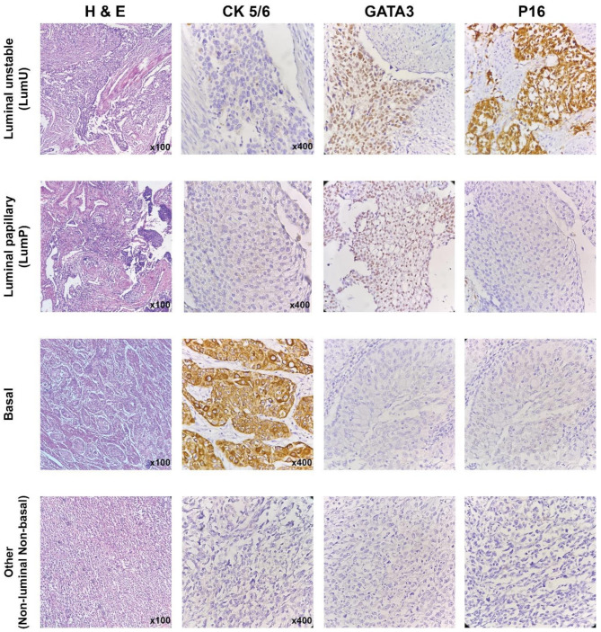
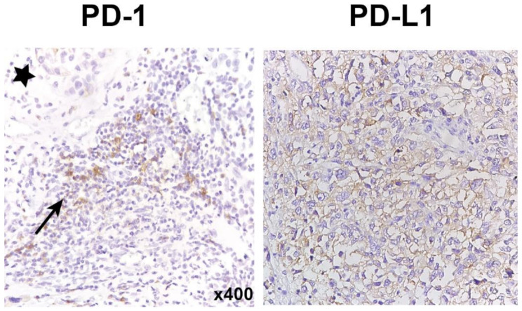
Methods: We evaluated 124 MIBC cases using IHC markers GATA3, CK5/6, and p16. Cases were classified as luminal (GATA3+, CK5/6-), basal (GATA3-, CK5/6+), or other (GATA3-, CK5/6-). Luminal cases were further subdivided into luminal unstable (LumU; p16+) and luminal papillary (LumP; p16-). Clinicopathological characteristics of MIBC molecular subtypes were also assessed. PD-1 and PD-L1 expression were evaluated relative to clinicopathological features and MIBC subtypes.
Results: In our study, 36.2% of the cases were LumU, 27.6% LumP, and 24.8% basal. The basal subtype generally shows a significantly higher tumor stage (p < 0.05). PD-1 was expressed in 70.5% of cases, with the highest expression in LumU (84.21%). PD-1 expression was significantly higher in the luminal compared to the basal subtype (82.1% vs. 53.8%, p < 0.01). PD-L1, expressed in 40% of cases, was significantly elevated in stage III and considerably higher in basal than luminal subtype (57.7% vs. 34.3%, p < 0.05).
Conclusion: MIBCs were practically subclassified into LumU, LumP, basal, and other subtypes using three IHC markers. PD-1 expression was higher in the luminal subtype, while PD-L1 was predominantly elevated in the basal subtype. These findings highlight the potential of IHC-based subtyping to guide prognosis and treatment in MIBCs.
期刊介绍:
Diagnostic Pathology is an open access, peer-reviewed, online journal that considers research in surgical and clinical pathology, immunology, and biology, with a special focus on cutting-edge approaches in diagnostic pathology and tissue-based therapy. The journal covers all aspects of surgical pathology, including classic diagnostic pathology, prognosis-related diagnosis (tumor stages, prognosis markers, such as MIB-percentage, hormone receptors, etc.), and therapy-related findings. The journal also focuses on the technological aspects of pathology, including molecular biology techniques, morphometry aspects (stereology, DNA analysis, syntactic structure analysis), communication aspects (telecommunication, virtual microscopy, virtual pathology institutions, etc.), and electronic education and quality assurance (for example interactive publication, on-line references with automated updating, etc.).

 求助内容:
求助内容: 应助结果提醒方式:


