Implications of imaging, morphologic, and immunohistochemical features of pancreatic perivascular epithelioid cell tumor: case report and comprehensive literature review.
Peipei He, Chaofeng Yang, Kexin Chen, Jinhong Yu, Yang Li
{"title":"Implications of imaging, morphologic, and immunohistochemical features of pancreatic perivascular epithelioid cell tumor: case report and comprehensive literature review.","authors":"Peipei He, Chaofeng Yang, Kexin Chen, Jinhong Yu, Yang Li","doi":"10.1186/s13000-025-01702-6","DOIUrl":null,"url":null,"abstract":"<p><strong>Background: </strong>Perivascular epithelioid cell tumor (PEComa) of the pancreas is a rare tumor of pancreatic mesenchymal origin with malignant potential. Critical to appropriate clinical management is determining whether the tumor is benign or malignant. Because of its rarity, morphologic and histologic characteristics and limited patient follow-up of pancreatic PEComa have precluded precise definition of malignancy. However, because malignant pancreatic PEComa appears to be distinctly uncommon, further improvements characterizing its preoperative imaging features could facilitate use of diagnostic endoscopic ultrasound biopsy and perhaps ablative treatment. This paper presents a case of pancreatic PEComa treated at the Affiliated Hospital of North Sichuan Medical College and includes a systematic literature review with special emphasis on the key imaging features of pancreatic PEComa.</p><p><strong>Case presentation: </strong>In February 2024, a woman in her 50s was admitted to the hospital with subxiphoid discomfort. Magnetic resonance imaging (MRI) of the upper abdomen revealed a round, solid mass in the pancreatic uncinate process. The patient underwent pancreatic mass resection and pancreaticojejunostomy, and the diagnosis of pancreatic PEComa was confirmed through pathological examination.</p><p><strong>Conclusions: </strong>Imaging examinations appear valuable for a tentative diagnosis of pancreatic PEComa. Key imaging features include its frequent occurrence in the pancreatic head, typically small to moderate size, \"pushing\" as opposed to infiltrative growth pattern with well-defined margins, and the presence of a capsule. The lesions are usually solid and often exhibit mild to moderate heterogenous enhancement during the arterial phase, with reduced enhancement in the portal and delayed phases.</p>","PeriodicalId":11237,"journal":{"name":"Diagnostic Pathology","volume":"20 1","pages":"95"},"PeriodicalIF":2.3000,"publicationDate":"2025-08-25","publicationTypes":"Journal Article","fieldsOfStudy":null,"isOpenAccess":false,"openAccessPdf":"https://www.ncbi.nlm.nih.gov/pmc/articles/PMC12376425/pdf/","citationCount":"0","resultStr":null,"platform":"Semanticscholar","paperid":null,"PeriodicalName":"Diagnostic Pathology","FirstCategoryId":"3","ListUrlMain":"https://doi.org/10.1186/s13000-025-01702-6","RegionNum":3,"RegionCategory":"医学","ArticlePicture":[],"TitleCN":null,"AbstractTextCN":null,"PMCID":null,"EPubDate":"","PubModel":"","JCR":"Q2","JCRName":"PATHOLOGY","Score":null,"Total":0}
引用次数: 0
Abstract
Background: Perivascular epithelioid cell tumor (PEComa) of the pancreas is a rare tumor of pancreatic mesenchymal origin with malignant potential. Critical to appropriate clinical management is determining whether the tumor is benign or malignant. Because of its rarity, morphologic and histologic characteristics and limited patient follow-up of pancreatic PEComa have precluded precise definition of malignancy. However, because malignant pancreatic PEComa appears to be distinctly uncommon, further improvements characterizing its preoperative imaging features could facilitate use of diagnostic endoscopic ultrasound biopsy and perhaps ablative treatment. This paper presents a case of pancreatic PEComa treated at the Affiliated Hospital of North Sichuan Medical College and includes a systematic literature review with special emphasis on the key imaging features of pancreatic PEComa.
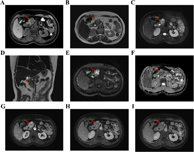
Case presentation: In February 2024, a woman in her 50s was admitted to the hospital with subxiphoid discomfort. Magnetic resonance imaging (MRI) of the upper abdomen revealed a round, solid mass in the pancreatic uncinate process. The patient underwent pancreatic mass resection and pancreaticojejunostomy, and the diagnosis of pancreatic PEComa was confirmed through pathological examination.
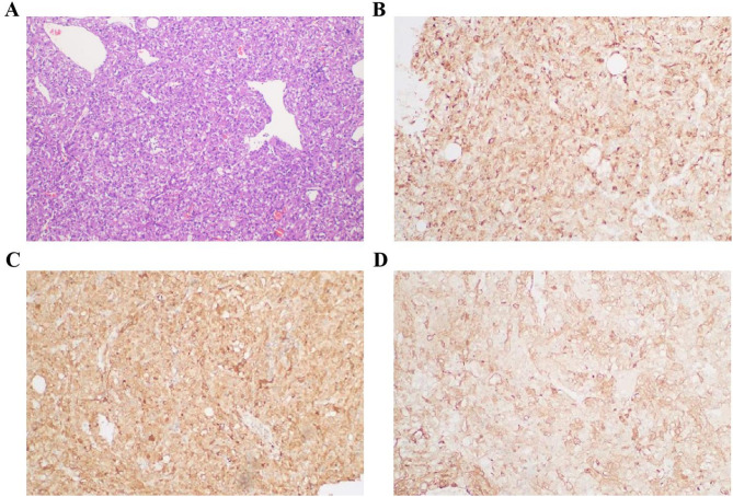
Conclusions: Imaging examinations appear valuable for a tentative diagnosis of pancreatic PEComa. Key imaging features include its frequent occurrence in the pancreatic head, typically small to moderate size, "pushing" as opposed to infiltrative growth pattern with well-defined margins, and the presence of a capsule. The lesions are usually solid and often exhibit mild to moderate heterogenous enhancement during the arterial phase, with reduced enhancement in the portal and delayed phases.
期刊介绍:
Diagnostic Pathology is an open access, peer-reviewed, online journal that considers research in surgical and clinical pathology, immunology, and biology, with a special focus on cutting-edge approaches in diagnostic pathology and tissue-based therapy. The journal covers all aspects of surgical pathology, including classic diagnostic pathology, prognosis-related diagnosis (tumor stages, prognosis markers, such as MIB-percentage, hormone receptors, etc.), and therapy-related findings. The journal also focuses on the technological aspects of pathology, including molecular biology techniques, morphometry aspects (stereology, DNA analysis, syntactic structure analysis), communication aspects (telecommunication, virtual microscopy, virtual pathology institutions, etc.), and electronic education and quality assurance (for example interactive publication, on-line references with automated updating, etc.).

 求助内容:
求助内容: 应助结果提醒方式:


