Bilateral, multicystic fumarate hydratase-deficient renal cell carcinoma in patient with hereditary leiomyomatosis & renal cell carcinoma syndrome: A case report and review of the literature.
Ashlie E Rubrecht, Jennifer H Aldrink, Patrick Warren, Mariam T Mathew, Karen Tsuchiya, Nicole Moulas, Vinay Prasad, Nilay Shah
{"title":"Bilateral, multicystic fumarate hydratase-deficient renal cell carcinoma in patient with hereditary leiomyomatosis & renal cell carcinoma syndrome: A case report and review of the literature.","authors":"Ashlie E Rubrecht, Jennifer H Aldrink, Patrick Warren, Mariam T Mathew, Karen Tsuchiya, Nicole Moulas, Vinay Prasad, Nilay Shah","doi":"10.1186/s13000-025-01706-2","DOIUrl":null,"url":null,"abstract":"<p><strong>Background: </strong>Hereditary leiomyomatosis and renal cell carcinoma syndrome (HLRCC) is an autosomal dominant tumor predisposition syndrome with germline fumarate hydratase (FH) pathogenic variants. We describe the unusual clinical presentation, morphologic, and immunohistochemical features of bilateral renal cell carcinoma (RCC) occurring in polycystic kidneys in a 15-year-old male with HLRCC.</p><p><strong>Case presentation: </strong>The patient was diagnosed with bilateral polycystic kidneys at 1-year old. At 8-years old he was diagnosed with cutaneous leiomyomas, prompting germline testing which revealed heterozygous variant (c.1301G > A) in the FH gene. Serial imaging identified interval enlargement of several bilateral renal lesions with solid components. Biopsy of a right solid lesion revealed an oncocytic neoplasm. He underwent left total nephrectomy and right partial nephrectomy, revealing numerous bilateral solid and cystic lesions, some with papillary excrescences. Histologic evaluation revealed large cells with eosinophilic to clear cytoplasm and large nuclei with occasional nuclear pseudoinclusions arranged in variable architectural patterns including papillary, tubular, tubulocystic, microcystic and solid. Large cysts were lined by varying thickness of neoplastic cells. By immunohistochemistry, lesional cells were positive for 2-succinocysteine (2SC), TFE3, PAX8 and AMACR, showed retained SDHB, variable FH, and were negative for Cathepsin K, CK20, and CK7. An RNA fusion panel (including TFE3) was negative. Multiple microscopic renal leiomyomas were also present.</p><p><strong>Conclusions: </strong>Multicystic kidney disease has been previously reported in HLRCC but is not currently included in the WHO classification. Bilateral involvement may mimic polycystic kidney disease and cysts may represent precursor lesions. TFE3-positivity raises the possibility of translocation RCC and is a diagnostic pitfall.</p>","PeriodicalId":11237,"journal":{"name":"Diagnostic Pathology","volume":"20 1","pages":"99"},"PeriodicalIF":2.3000,"publicationDate":"2025-08-26","publicationTypes":"Journal Article","fieldsOfStudy":null,"isOpenAccess":false,"openAccessPdf":"https://www.ncbi.nlm.nih.gov/pmc/articles/PMC12379367/pdf/","citationCount":"0","resultStr":null,"platform":"Semanticscholar","paperid":null,"PeriodicalName":"Diagnostic Pathology","FirstCategoryId":"3","ListUrlMain":"https://doi.org/10.1186/s13000-025-01706-2","RegionNum":3,"RegionCategory":"医学","ArticlePicture":[],"TitleCN":null,"AbstractTextCN":null,"PMCID":null,"EPubDate":"","PubModel":"","JCR":"Q2","JCRName":"PATHOLOGY","Score":null,"Total":0}
引用次数: 0
Abstract
Background: Hereditary leiomyomatosis and renal cell carcinoma syndrome (HLRCC) is an autosomal dominant tumor predisposition syndrome with germline fumarate hydratase (FH) pathogenic variants. We describe the unusual clinical presentation, morphologic, and immunohistochemical features of bilateral renal cell carcinoma (RCC) occurring in polycystic kidneys in a 15-year-old male with HLRCC.
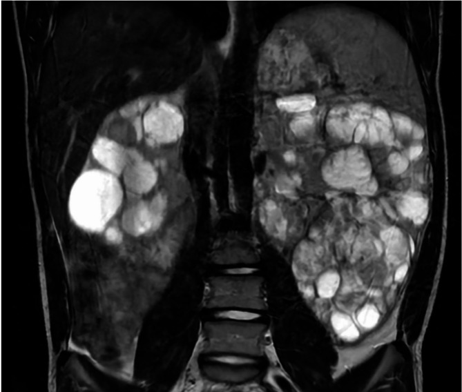
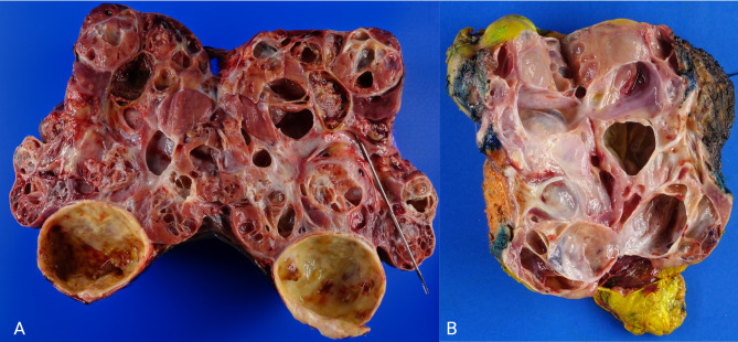
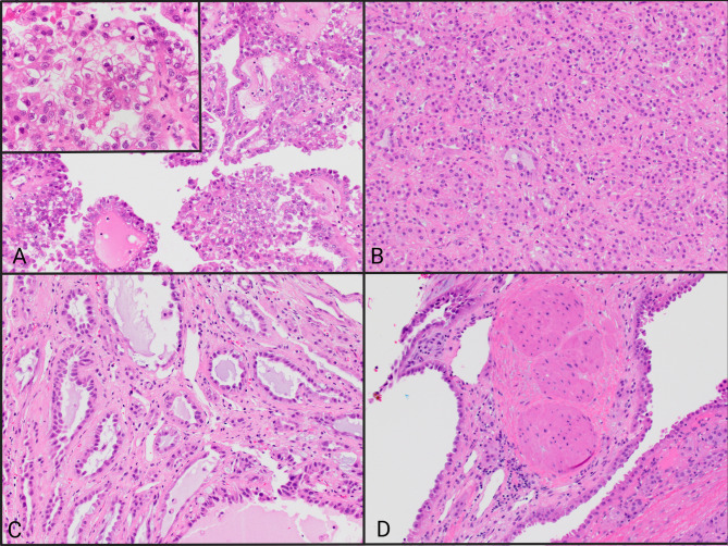
Case presentation: The patient was diagnosed with bilateral polycystic kidneys at 1-year old. At 8-years old he was diagnosed with cutaneous leiomyomas, prompting germline testing which revealed heterozygous variant (c.1301G > A) in the FH gene. Serial imaging identified interval enlargement of several bilateral renal lesions with solid components. Biopsy of a right solid lesion revealed an oncocytic neoplasm. He underwent left total nephrectomy and right partial nephrectomy, revealing numerous bilateral solid and cystic lesions, some with papillary excrescences. Histologic evaluation revealed large cells with eosinophilic to clear cytoplasm and large nuclei with occasional nuclear pseudoinclusions arranged in variable architectural patterns including papillary, tubular, tubulocystic, microcystic and solid. Large cysts were lined by varying thickness of neoplastic cells. By immunohistochemistry, lesional cells were positive for 2-succinocysteine (2SC), TFE3, PAX8 and AMACR, showed retained SDHB, variable FH, and were negative for Cathepsin K, CK20, and CK7. An RNA fusion panel (including TFE3) was negative. Multiple microscopic renal leiomyomas were also present.
Conclusions: Multicystic kidney disease has been previously reported in HLRCC but is not currently included in the WHO classification. Bilateral involvement may mimic polycystic kidney disease and cysts may represent precursor lesions. TFE3-positivity raises the possibility of translocation RCC and is a diagnostic pitfall.
期刊介绍:
Diagnostic Pathology is an open access, peer-reviewed, online journal that considers research in surgical and clinical pathology, immunology, and biology, with a special focus on cutting-edge approaches in diagnostic pathology and tissue-based therapy. The journal covers all aspects of surgical pathology, including classic diagnostic pathology, prognosis-related diagnosis (tumor stages, prognosis markers, such as MIB-percentage, hormone receptors, etc.), and therapy-related findings. The journal also focuses on the technological aspects of pathology, including molecular biology techniques, morphometry aspects (stereology, DNA analysis, syntactic structure analysis), communication aspects (telecommunication, virtual microscopy, virtual pathology institutions, etc.), and electronic education and quality assurance (for example interactive publication, on-line references with automated updating, etc.).

 求助内容:
求助内容: 应助结果提醒方式:


