{"title":"Evaluation of Compatibility of Different Attachment Types Used in Orthodontic Clear Aligners with Electron Microscopy.","authors":"Can Sever, Can Arslan","doi":"10.3390/dj13080379","DOIUrl":null,"url":null,"abstract":"<p><p><b>Background/Objectives</b>: The effectiveness of clear aligner therapy depends significantly on the precision of force delivery through the aligner-attachment interface. This study aimed to evaluate the microscopic compatibility between different orthodontic clear aligner materials (Duran+ and Zendura FLX) and attachment designs (rectangular and optimized) using scanning electron microscopy (SEM). <b>Methods</b>: Fifty-six samples were divided into four groups: rectangular attachments with Duran+ aligners (n = 14), rectangular attachments with Zendura FLX aligners (n = 14), optimized attachments with Duran+ aligners (n = 14), and optimized attachments with Zendura FLX aligners (n = 14). Attachments were bonded to bovine incisors using standardized protocols. Clear aligners were thermoformed at 220 °C for 40 s. Cross-sectional samples were analyzed using SEM at 250× magnification. Gap measurements were taken at seven points for rectangular attachments and five points for optimized attachments. <b>Results</b>: Gap measurements ranged from 14.75 ± 1.41 µm to 91.07 ± 3.11 µm. Zendura FLX demonstrated significantly better adaptation than Duran+ with rectangular attachments (42.10 ± 1.07 µm vs. 44.52 ± 1.51 µm, <i>p</i> < 0.001). Optimized attachments showed better overall adaptation than rectangular attachments. All combinations showed regional variation with the largest gaps at gingival borders (67.18-91.07 µm) and the smallest at flat buccal surfaces (14.75-20.98 µm). <b>Conclusions</b>: Perfect adaptation was not achieved with any material-attachment combination tested. Material selection and attachment design significantly influence microscopic adaptation, with multi-layer materials and optimized geometries showing superior performance. These findings provide mechanical explanations for clinical limitations in clear aligner therapy.</p>","PeriodicalId":11269,"journal":{"name":"Dentistry Journal","volume":"13 8","pages":""},"PeriodicalIF":3.1000,"publicationDate":"2025-08-20","publicationTypes":"Journal Article","fieldsOfStudy":null,"isOpenAccess":false,"openAccessPdf":"https://www.ncbi.nlm.nih.gov/pmc/articles/PMC12385661/pdf/","citationCount":"0","resultStr":null,"platform":"Semanticscholar","paperid":null,"PeriodicalName":"Dentistry Journal","FirstCategoryId":"1085","ListUrlMain":"https://doi.org/10.3390/dj13080379","RegionNum":0,"RegionCategory":null,"ArticlePicture":[],"TitleCN":null,"AbstractTextCN":null,"PMCID":null,"EPubDate":"","PubModel":"","JCR":"Q2","JCRName":"DENTISTRY, ORAL SURGERY & MEDICINE","Score":null,"Total":0}
引用次数: 0
Abstract
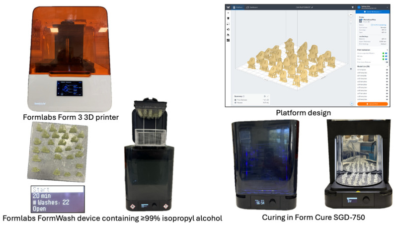
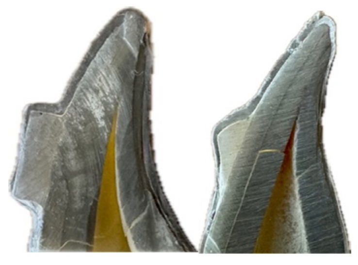
Background/Objectives: The effectiveness of clear aligner therapy depends significantly on the precision of force delivery through the aligner-attachment interface. This study aimed to evaluate the microscopic compatibility between different orthodontic clear aligner materials (Duran+ and Zendura FLX) and attachment designs (rectangular and optimized) using scanning electron microscopy (SEM). Methods: Fifty-six samples were divided into four groups: rectangular attachments with Duran+ aligners (n = 14), rectangular attachments with Zendura FLX aligners (n = 14), optimized attachments with Duran+ aligners (n = 14), and optimized attachments with Zendura FLX aligners (n = 14). Attachments were bonded to bovine incisors using standardized protocols. Clear aligners were thermoformed at 220 °C for 40 s. Cross-sectional samples were analyzed using SEM at 250× magnification. Gap measurements were taken at seven points for rectangular attachments and five points for optimized attachments. Results: Gap measurements ranged from 14.75 ± 1.41 µm to 91.07 ± 3.11 µm. Zendura FLX demonstrated significantly better adaptation than Duran+ with rectangular attachments (42.10 ± 1.07 µm vs. 44.52 ± 1.51 µm, p < 0.001). Optimized attachments showed better overall adaptation than rectangular attachments. All combinations showed regional variation with the largest gaps at gingival borders (67.18-91.07 µm) and the smallest at flat buccal surfaces (14.75-20.98 µm). Conclusions: Perfect adaptation was not achieved with any material-attachment combination tested. Material selection and attachment design significantly influence microscopic adaptation, with multi-layer materials and optimized geometries showing superior performance. These findings provide mechanical explanations for clinical limitations in clear aligner therapy.

 求助内容:
求助内容: 应助结果提醒方式:


