{"title":"Urinary microvesicles: a window into the kidney.","authors":"Luisa Schnobrich, Hayo Castrop","doi":"10.1093/ckj/sfaf189","DOIUrl":null,"url":null,"abstract":"<p><p>Chronic kidney disease (CKD) is a growing concern in aging populations. CKD is characterized by two hallmark symptoms: a decline in the glomerular filtration rate (GFR) and albuminuria. Early changes in kidney function are notoriously underdiagnosed, suggesting the need for new noninvasive diagnostic and prognostic biomarkers of CKD. Thus, analysis of urinary extracellular vesicles (uEVs) may broaden the diagnostic options for CKD. EVs are a heterogeneous group of particles, enclosed by a lipid bilayer, which differ in size, biogenesis, and function. EVs can be readily recovered from the urine (urinary EVs, uEVs), where they are derived from various cells of the kidney, bladder, prostate, and utero-vaginal tract. Within the kidney, EVs are released by almost all cell types, including but not limited to podocytes, cells of the proximal and distal tubules, the collecting duct, and the loop of Henle. In addition to specific markers of parental cells, uEVs carry mRNAs, miRNAs, and proteins. Thus, analysis of uEVs may provide insights into the content and composition of the specific cells from which they are released, leading to the identification of new diagnostic and prognostic biomarkers for kidney diseases of different etiologies. This review provides an overview of kidney disease-related changes in uEV size and concentration and covers the potential of uEVs as new biomarkers for various types of kidney disease.</p>","PeriodicalId":10435,"journal":{"name":"Clinical Kidney Journal","volume":"18 7","pages":"sfaf189"},"PeriodicalIF":4.6000,"publicationDate":"2025-06-17","publicationTypes":"Journal Article","fieldsOfStudy":null,"isOpenAccess":false,"openAccessPdf":"https://www.ncbi.nlm.nih.gov/pmc/articles/PMC12378445/pdf/","citationCount":"0","resultStr":null,"platform":"Semanticscholar","paperid":null,"PeriodicalName":"Clinical Kidney Journal","FirstCategoryId":"3","ListUrlMain":"https://doi.org/10.1093/ckj/sfaf189","RegionNum":2,"RegionCategory":"医学","ArticlePicture":[],"TitleCN":null,"AbstractTextCN":null,"PMCID":null,"EPubDate":"2025/7/1 0:00:00","PubModel":"eCollection","JCR":"Q1","JCRName":"UROLOGY & NEPHROLOGY","Score":null,"Total":0}
引用次数: 0
Abstract
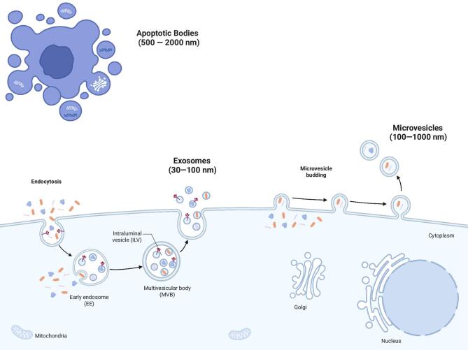
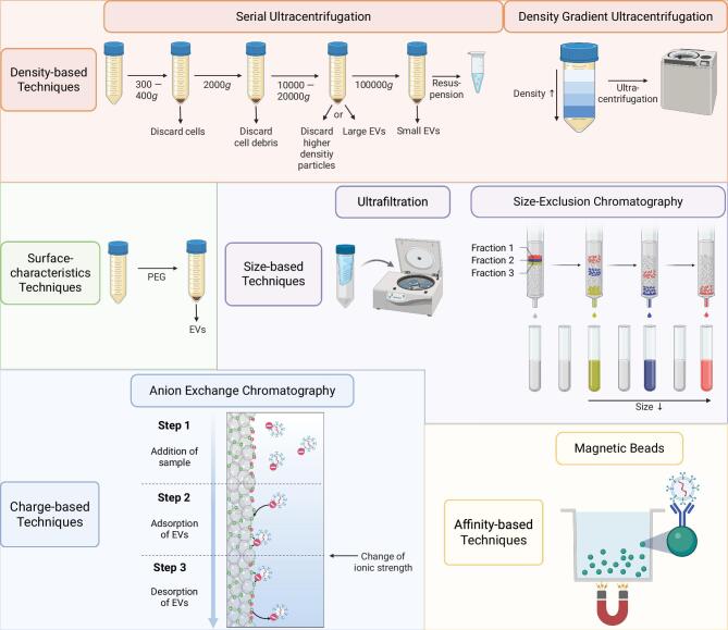
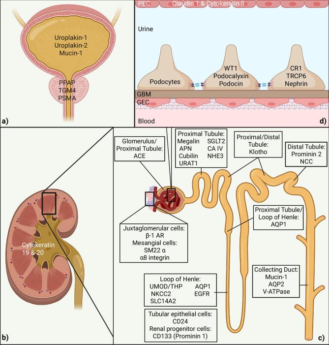
Chronic kidney disease (CKD) is a growing concern in aging populations. CKD is characterized by two hallmark symptoms: a decline in the glomerular filtration rate (GFR) and albuminuria. Early changes in kidney function are notoriously underdiagnosed, suggesting the need for new noninvasive diagnostic and prognostic biomarkers of CKD. Thus, analysis of urinary extracellular vesicles (uEVs) may broaden the diagnostic options for CKD. EVs are a heterogeneous group of particles, enclosed by a lipid bilayer, which differ in size, biogenesis, and function. EVs can be readily recovered from the urine (urinary EVs, uEVs), where they are derived from various cells of the kidney, bladder, prostate, and utero-vaginal tract. Within the kidney, EVs are released by almost all cell types, including but not limited to podocytes, cells of the proximal and distal tubules, the collecting duct, and the loop of Henle. In addition to specific markers of parental cells, uEVs carry mRNAs, miRNAs, and proteins. Thus, analysis of uEVs may provide insights into the content and composition of the specific cells from which they are released, leading to the identification of new diagnostic and prognostic biomarkers for kidney diseases of different etiologies. This review provides an overview of kidney disease-related changes in uEV size and concentration and covers the potential of uEVs as new biomarkers for various types of kidney disease.
期刊介绍:
About the Journal
Clinical Kidney Journal: Clinical and Translational Nephrology (ckj), an official journal of the ERA-EDTA (European Renal Association-European Dialysis and Transplant Association), is a fully open access, online only journal publishing bimonthly. The journal is an essential educational and training resource integrating clinical, translational and educational research into clinical practice. ckj aims to contribute to a translational research culture among nephrologists and kidney pathologists that helps close the gap between basic researchers and practicing clinicians and promote sorely needed innovation in the Nephrology field. All research articles in this journal have undergone peer review.
 求助内容:
求助内容: 应助结果提醒方式:


