Zouheir Ibrahim Bitar, Ossama Sajeh Maadarani, Mohamad Abdelfatah, Bibin Antony
{"title":"Pneumatosis Intestinalis Detected by Point of Care Ultrasound, Case Report.","authors":"Zouheir Ibrahim Bitar, Ossama Sajeh Maadarani, Mohamad Abdelfatah, Bibin Antony","doi":"10.1177/11795476251370547","DOIUrl":null,"url":null,"abstract":"<p><p>Pneumatosis intestinalis (PI) is characterized by the presence of air within the walls of the small intestine, large intestine, and sometimes the gastric wall. The mechanism and pathogenesis of PI are poorly understood. The discovery of PI can occur in the form of an incidental finding, such as a benign course or a life-threatening condition, such as intestinal ischemia. Point-of-care ultrasonography (POCUS) for pneumatosis intestinalis (PI) is rarely reported in adults, with only 1 previous poster presentation. However, POCUS is well-documented in the pediatric population, particularly for the diagnosis of intussusception. We present a 78-year-old man with longstanding uncorrected severe pulmonary stenosis, right-sided heart failure, atrial fibrillation on dabigatran, diabetes, and cirrhosis who presented with progressive abdominal distension. POCUS and computerized tomography of the abdomen showed ascites, diffusing small-bowel wall thickening with edema, and pneumatosis intestinalis, without portal venous gas or vascular occlusion. Patient was transitioned to palliative care on diuretics, lactulose; follow-up ultrasound showed only mild ascites but persistent small-bowel PI. This case report marks the second instance of POCUS being utilized for PI in an adult population. POCUS can play an essential diagnostic role at the bedside, ruling out serious underlying etiologies and guiding physicians in further diagnostic testing.</p>","PeriodicalId":10357,"journal":{"name":"Clinical Medicine Insights. Case Reports","volume":"18 ","pages":"11795476251370547"},"PeriodicalIF":0.6000,"publicationDate":"2025-08-29","publicationTypes":"Journal Article","fieldsOfStudy":null,"isOpenAccess":false,"openAccessPdf":"https://www.ncbi.nlm.nih.gov/pmc/articles/PMC12397599/pdf/","citationCount":"0","resultStr":null,"platform":"Semanticscholar","paperid":null,"PeriodicalName":"Clinical Medicine Insights. Case Reports","FirstCategoryId":"1085","ListUrlMain":"https://doi.org/10.1177/11795476251370547","RegionNum":0,"RegionCategory":null,"ArticlePicture":[],"TitleCN":null,"AbstractTextCN":null,"PMCID":null,"EPubDate":"2025/1/1 0:00:00","PubModel":"eCollection","JCR":"Q3","JCRName":"MEDICINE, GENERAL & INTERNAL","Score":null,"Total":0}
引用次数: 0
Abstract
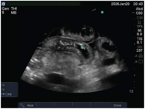
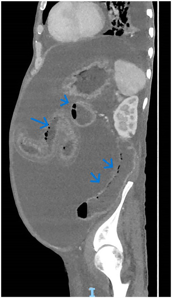
Pneumatosis intestinalis (PI) is characterized by the presence of air within the walls of the small intestine, large intestine, and sometimes the gastric wall. The mechanism and pathogenesis of PI are poorly understood. The discovery of PI can occur in the form of an incidental finding, such as a benign course or a life-threatening condition, such as intestinal ischemia. Point-of-care ultrasonography (POCUS) for pneumatosis intestinalis (PI) is rarely reported in adults, with only 1 previous poster presentation. However, POCUS is well-documented in the pediatric population, particularly for the diagnosis of intussusception. We present a 78-year-old man with longstanding uncorrected severe pulmonary stenosis, right-sided heart failure, atrial fibrillation on dabigatran, diabetes, and cirrhosis who presented with progressive abdominal distension. POCUS and computerized tomography of the abdomen showed ascites, diffusing small-bowel wall thickening with edema, and pneumatosis intestinalis, without portal venous gas or vascular occlusion. Patient was transitioned to palliative care on diuretics, lactulose; follow-up ultrasound showed only mild ascites but persistent small-bowel PI. This case report marks the second instance of POCUS being utilized for PI in an adult population. POCUS can play an essential diagnostic role at the bedside, ruling out serious underlying etiologies and guiding physicians in further diagnostic testing.


 求助内容:
求助内容: 应助结果提醒方式:


