Contrast-enhanced ultrasound for diagnosing subtypes of intrahepatic cholangiocarcinoma: a comparative study with poorly differentiated hepatocellular carcinoma.
Nan Zhang, Yue Yang, Ke Lin, Bin Qiao, Dao-Peng Yang, Dong-Dong Jin, Bin Li, Dong-Liang Zhao, Xiao-Hua Xie, Xiao-Yan Xie, Ji-Hui Kang, Bo-Wen Zhuang
{"title":"Contrast-enhanced ultrasound for diagnosing subtypes of intrahepatic cholangiocarcinoma: a comparative study with poorly differentiated hepatocellular carcinoma.","authors":"Nan Zhang, Yue Yang, Ke Lin, Bin Qiao, Dao-Peng Yang, Dong-Dong Jin, Bin Li, Dong-Liang Zhao, Xiao-Hua Xie, Xiao-Yan Xie, Ji-Hui Kang, Bo-Wen Zhuang","doi":"10.1186/s40644-025-00923-8","DOIUrl":null,"url":null,"abstract":"<p><strong>Background: </strong>Pathologically, intrahepatic cholangiocarcinoma (ICC) is classified into small-duct (SD) type and large-duct (LD) type, each with distinct clinicopathological characteristics. The contrast-enhanced ultrasound (CEUS) features of the two ICC types remain insufficiently explored.</p><p><strong>Purpose: </strong>To evaluate liver CEUS imaging for differentiating the SD and LD types of ICC and further compare them with poorly differentiated hepatocellular carcinoma (pHCC).</p><p><strong>Materials and methods: </strong>A single-center retrospective study enrolled 252 patients with SD-type ICC, LD-type ICC, or pHCC between October 2017 and August 2023. Logistic regression analyses identified independent clinical, pathological, ultrasound, and CEUS predictors. Based on these features, a decision tree-based diagnostic model was developed. The model's performance was evaluated using receiver operating characteristic (ROC) curve analysis in both the training and validation cohorts, as well as in subgroup stratified by tumor size ≤ 5 cm and > 5 cm. Differences in overall survival (OS) and recurrence-free survival (RFS) based on the model were further analyzed.</p><p><strong>Results: </strong>Overall, 252 patients (mean age, 58.4 ± 10.7 years; 174 males) with 140 SD-type ICC, 55 LD-type ICC and 57 pHCC were enrolled. Multivariate analysis revealed that AFP, CEA, CA19-9, HBsAg status, arterial phase enhancement pattern, washout time ≤ 45 s, and marked washout were independent predictors for tumor categories differentiation (all P <.05). The decision tree-based model incorporating the major features demonstrated excellent performance in both the training cohort (AUC 0.89) and validation cohort (AUC 0.88), as well as in tumor size ≤ 5 cm (AUC 0.90) and > 5 cm (AUC 0.84). OS was significantly worse in LD-type ICC patients compared to SD-type and pHCC (P <.05 for both), while RFS showed no significant difference.</p><p><strong>Conclusions: </strong>A user-friendly, decision tree-based diagnostic model was developed to accurately predict ICC subtypes and pHCC, facilitating improved clinical decision-making. The decision tree-based diagnostic model effectively diagnosed small-duct type and large-duct type intrahepatic cholangiocarcinoma, as well as poorly differentiated hepatocellular carcinoma.</p>","PeriodicalId":9548,"journal":{"name":"Cancer Imaging","volume":"25 1","pages":"107"},"PeriodicalIF":3.5000,"publicationDate":"2025-08-27","publicationTypes":"Journal Article","fieldsOfStudy":null,"isOpenAccess":false,"openAccessPdf":"https://www.ncbi.nlm.nih.gov/pmc/articles/PMC12382020/pdf/","citationCount":"0","resultStr":null,"platform":"Semanticscholar","paperid":null,"PeriodicalName":"Cancer Imaging","FirstCategoryId":"3","ListUrlMain":"https://doi.org/10.1186/s40644-025-00923-8","RegionNum":2,"RegionCategory":"医学","ArticlePicture":[],"TitleCN":null,"AbstractTextCN":null,"PMCID":null,"EPubDate":"","PubModel":"","JCR":"Q2","JCRName":"ONCOLOGY","Score":null,"Total":0}
引用次数: 0
Abstract
Background: Pathologically, intrahepatic cholangiocarcinoma (ICC) is classified into small-duct (SD) type and large-duct (LD) type, each with distinct clinicopathological characteristics. The contrast-enhanced ultrasound (CEUS) features of the two ICC types remain insufficiently explored.
Purpose: To evaluate liver CEUS imaging for differentiating the SD and LD types of ICC and further compare them with poorly differentiated hepatocellular carcinoma (pHCC).
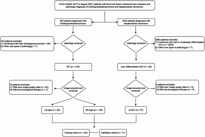
Materials and methods: A single-center retrospective study enrolled 252 patients with SD-type ICC, LD-type ICC, or pHCC between October 2017 and August 2023. Logistic regression analyses identified independent clinical, pathological, ultrasound, and CEUS predictors. Based on these features, a decision tree-based diagnostic model was developed. The model's performance was evaluated using receiver operating characteristic (ROC) curve analysis in both the training and validation cohorts, as well as in subgroup stratified by tumor size ≤ 5 cm and > 5 cm. Differences in overall survival (OS) and recurrence-free survival (RFS) based on the model were further analyzed.
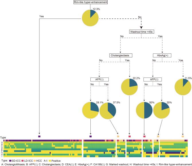
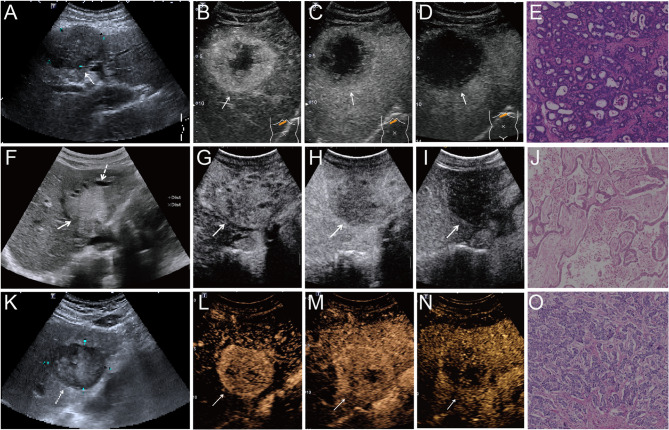
Results: Overall, 252 patients (mean age, 58.4 ± 10.7 years; 174 males) with 140 SD-type ICC, 55 LD-type ICC and 57 pHCC were enrolled. Multivariate analysis revealed that AFP, CEA, CA19-9, HBsAg status, arterial phase enhancement pattern, washout time ≤ 45 s, and marked washout were independent predictors for tumor categories differentiation (all P <.05). The decision tree-based model incorporating the major features demonstrated excellent performance in both the training cohort (AUC 0.89) and validation cohort (AUC 0.88), as well as in tumor size ≤ 5 cm (AUC 0.90) and > 5 cm (AUC 0.84). OS was significantly worse in LD-type ICC patients compared to SD-type and pHCC (P <.05 for both), while RFS showed no significant difference.
Conclusions: A user-friendly, decision tree-based diagnostic model was developed to accurately predict ICC subtypes and pHCC, facilitating improved clinical decision-making. The decision tree-based diagnostic model effectively diagnosed small-duct type and large-duct type intrahepatic cholangiocarcinoma, as well as poorly differentiated hepatocellular carcinoma.
Cancer ImagingONCOLOGY-RADIOLOGY, NUCLEAR MEDICINE & MEDICAL IMAGING
CiteScore
7.00
自引率
0.00%
发文量
66
审稿时长
>12 weeks
期刊介绍:
Cancer Imaging is an open access, peer-reviewed journal publishing original articles, reviews and editorials written by expert international radiologists working in oncology.
The journal encompasses CT, MR, PET, ultrasound, radionuclide and multimodal imaging in all kinds of malignant tumours, plus new developments, techniques and innovations. Topics of interest include:
Breast Imaging
Chest
Complications of treatment
Ear, Nose & Throat
Gastrointestinal
Hepatobiliary & Pancreatic
Imaging biomarkers
Interventional
Lymphoma
Measurement of tumour response
Molecular functional imaging
Musculoskeletal
Neuro oncology
Nuclear Medicine
Paediatric.

 求助内容:
求助内容: 应助结果提醒方式:


