Ali Shahriari, Sasan Ghazanafar Ahari, Ali Mousavi, Mahdie Sadeghi, Marjan Abbasi, Mahsa Hosseinpour, Asal Mir, Dorrin Zohouri Zanganeh, Hossein Gharedaghi, Saba Ezati, Ali Sareminia, Dina Seyedi, Mahla Shokouhfar, Ali Darzi, Alireza Ghaedamini, Sara Zamani, Farbod Khosravi, Mahsa Asadi Anar
{"title":"Machine Learning-Driven radiomics on 18 F-FDG PET for glioma diagnosis: a systematic review and meta-analysis.","authors":"Ali Shahriari, Sasan Ghazanafar Ahari, Ali Mousavi, Mahdie Sadeghi, Marjan Abbasi, Mahsa Hosseinpour, Asal Mir, Dorrin Zohouri Zanganeh, Hossein Gharedaghi, Saba Ezati, Ali Sareminia, Dina Seyedi, Mahla Shokouhfar, Ali Darzi, Alireza Ghaedamini, Sara Zamani, Farbod Khosravi, Mahsa Asadi Anar","doi":"10.1186/s40644-025-00915-8","DOIUrl":null,"url":null,"abstract":"<p><strong>Background: </strong>Machine learning (ML) applied to radiomics has revolutionized neuro-oncological imaging, yet the diagnostic performance of ML models based specifically on ^18F-FDG PET features in glioma remains poorly characterized.</p><p><strong>Objective: </strong>To systematically evaluate and quantitatively synthesize the diagnostic accuracy of ML models trained on ^18F-FDG PET radiomics for glioma classification.</p><p><strong>Methods: </strong>We conducted a PRISMA-compliant systematic review and meta-analysis registered on OSF ( https://doi.org/10.17605/OSF.IO/XJG6P ). PubMed, Scopus, and Web of Science were searched up to January 2025. Studies were included if they applied ML algorithms to ^18F-FDG PET radiomic features for glioma classification and reported at least one performance metric. Data extraction included demographics, imaging protocols, feature types, ML models, and validation design. Meta-analysis was performed using random-effects models with pooled estimates of accuracy, sensitivity, specificity, AUC, F1 score, and precision. Heterogeneity was explored via meta-regression and Galbraith plots.</p><p><strong>Results: </strong>Twelve studies comprising 2,321 patients were included. Pooled diagnostic metrics were: accuracy 92.6% (95% CI: 91.3-93.9%), AUC 0.95 (95% CI: 0.94-0.95), sensitivity 85.4%, specificity 89.7%, F1 score 0.78, and precision 0.90. Heterogeneity was high across all domains (I² >75%). Meta-regression identified ML model type and validation strategy as partial moderators. Models using CNNs or PET/MRI integration achieved superior performance.</p><p><strong>Conclusion: </strong>ML models based on ^18F-FDG PET radiomics demonstrate strong and balanced diagnostic performance for glioma classification. However, methodological heterogeneity underscores the need for standardized pipelines, external validation, and transparent reporting before clinical integration.</p>","PeriodicalId":9548,"journal":{"name":"Cancer Imaging","volume":"25 1","pages":"106"},"PeriodicalIF":3.5000,"publicationDate":"2025-08-26","publicationTypes":"Journal Article","fieldsOfStudy":null,"isOpenAccess":false,"openAccessPdf":"https://www.ncbi.nlm.nih.gov/pmc/articles/PMC12379540/pdf/","citationCount":"0","resultStr":null,"platform":"Semanticscholar","paperid":null,"PeriodicalName":"Cancer Imaging","FirstCategoryId":"3","ListUrlMain":"https://doi.org/10.1186/s40644-025-00915-8","RegionNum":2,"RegionCategory":"医学","ArticlePicture":[],"TitleCN":null,"AbstractTextCN":null,"PMCID":null,"EPubDate":"","PubModel":"","JCR":"Q2","JCRName":"ONCOLOGY","Score":null,"Total":0}
引用次数: 0
Abstract
Background: Machine learning (ML) applied to radiomics has revolutionized neuro-oncological imaging, yet the diagnostic performance of ML models based specifically on ^18F-FDG PET features in glioma remains poorly characterized.
Objective: To systematically evaluate and quantitatively synthesize the diagnostic accuracy of ML models trained on ^18F-FDG PET radiomics for glioma classification.
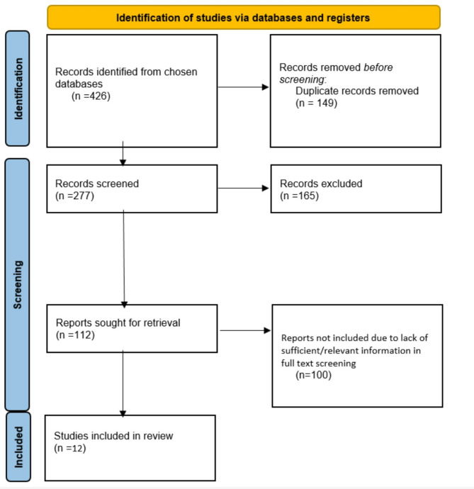
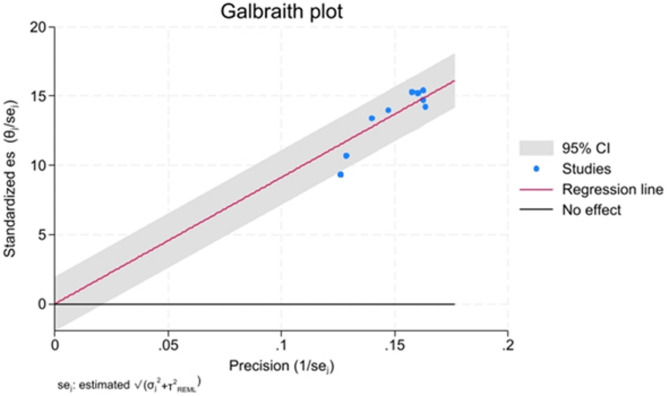
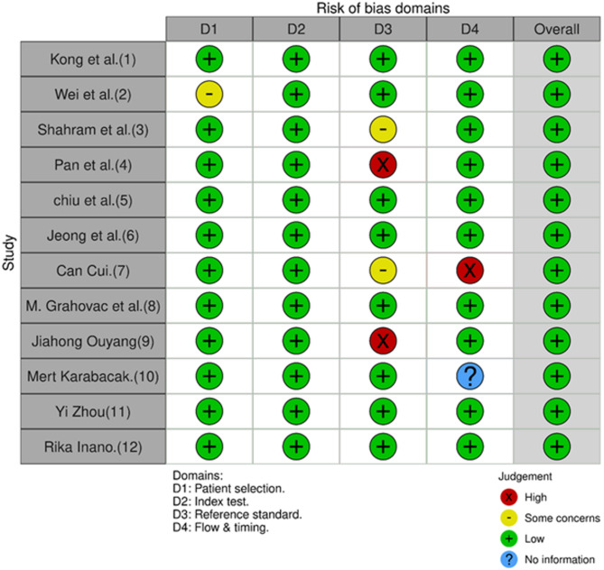
Methods: We conducted a PRISMA-compliant systematic review and meta-analysis registered on OSF ( https://doi.org/10.17605/OSF.IO/XJG6P ). PubMed, Scopus, and Web of Science were searched up to January 2025. Studies were included if they applied ML algorithms to ^18F-FDG PET radiomic features for glioma classification and reported at least one performance metric. Data extraction included demographics, imaging protocols, feature types, ML models, and validation design. Meta-analysis was performed using random-effects models with pooled estimates of accuracy, sensitivity, specificity, AUC, F1 score, and precision. Heterogeneity was explored via meta-regression and Galbraith plots.
Results: Twelve studies comprising 2,321 patients were included. Pooled diagnostic metrics were: accuracy 92.6% (95% CI: 91.3-93.9%), AUC 0.95 (95% CI: 0.94-0.95), sensitivity 85.4%, specificity 89.7%, F1 score 0.78, and precision 0.90. Heterogeneity was high across all domains (I² >75%). Meta-regression identified ML model type and validation strategy as partial moderators. Models using CNNs or PET/MRI integration achieved superior performance.
Conclusion: ML models based on ^18F-FDG PET radiomics demonstrate strong and balanced diagnostic performance for glioma classification. However, methodological heterogeneity underscores the need for standardized pipelines, external validation, and transparent reporting before clinical integration.
Cancer ImagingONCOLOGY-RADIOLOGY, NUCLEAR MEDICINE & MEDICAL IMAGING
CiteScore
7.00
自引率
0.00%
发文量
66
审稿时长
>12 weeks
期刊介绍:
Cancer Imaging is an open access, peer-reviewed journal publishing original articles, reviews and editorials written by expert international radiologists working in oncology.
The journal encompasses CT, MR, PET, ultrasound, radionuclide and multimodal imaging in all kinds of malignant tumours, plus new developments, techniques and innovations. Topics of interest include:
Breast Imaging
Chest
Complications of treatment
Ear, Nose & Throat
Gastrointestinal
Hepatobiliary & Pancreatic
Imaging biomarkers
Interventional
Lymphoma
Measurement of tumour response
Molecular functional imaging
Musculoskeletal
Neuro oncology
Nuclear Medicine
Paediatric.

 求助内容:
求助内容: 应助结果提醒方式:


