Jitong Zhou, Yuxi Chen, Jiaqi Meng, Kaiwen Cheng, Jiao Qi, Yu Du, Yunqian Yao, Yi Lu, Wenwen He, Xiangjia Zhu
{"title":"Comparative analysis of iris blood flow after first-eye and second-eye cataract surgeries: insights into increased pain perception in the second eye.","authors":"Jitong Zhou, Yuxi Chen, Jiaqi Meng, Kaiwen Cheng, Jiao Qi, Yu Du, Yunqian Yao, Yi Lu, Wenwen He, Xiangjia Zhu","doi":"10.1136/bmjophth-2025-002162","DOIUrl":null,"url":null,"abstract":"<p><strong>Objective: </strong>To investigate quantitative changes in iris blood flow after first-eye and second-eye cataract surgeries, and their correlation with increased pain in the second eye.</p><p><strong>Methods and analysis: </strong>In this prospective study, 88 eyes of 44 participants who underwent uneventful cataract surgery were followed up at enrolment, 1 day before, and 1 day, 1 week and 1 month after each eye surgery. Iris blood flow was quantified by a swept-source optical coherence tomography angiography and intraoperative pain was evaluated. Participants were divided into three groups according to time intervals between two eye surgeries: ≥1 to <2 weeks (short interval), ≥2 to ≤4 weeks (medium interval) and greater than 4 weeks (long interval).</p><p><strong>Results: </strong>The second eye experienced two significant increases in iris blood flow following cataract surgery: one after the first eye and the other after its own, with the latter being significantly higher than the increase observed in the operated eye after the first surgery (p<0.05). Additionally, the second eye showed higher iris blood flow density in the short interval group compared with the long and medium at specific time points (p<0.05). The bilateral pain index difference positively correlated with second eye iris blood flow density (p<0.05) and was significantly greater in the short interval group than the long interval group (p<0.05).</p><p><strong>Conclusions: </strong>Iris blood flow increased in the second-operated eye post surgery compared with the first eye, which may correlate to second-eye pain. However, due to the variability in individual pain perception, a larger sample size is needed to prospectively validate our findings to improve their generalisability.</p><p><strong>Trial registration number: </strong>NCT02182921.</p>","PeriodicalId":9286,"journal":{"name":"BMJ Open Ophthalmology","volume":"10 1","pages":""},"PeriodicalIF":2.2000,"publicationDate":"2025-08-19","publicationTypes":"Journal Article","fieldsOfStudy":null,"isOpenAccess":false,"openAccessPdf":"https://www.ncbi.nlm.nih.gov/pmc/articles/PMC12374657/pdf/","citationCount":"0","resultStr":null,"platform":"Semanticscholar","paperid":null,"PeriodicalName":"BMJ Open Ophthalmology","FirstCategoryId":"1085","ListUrlMain":"https://doi.org/10.1136/bmjophth-2025-002162","RegionNum":0,"RegionCategory":null,"ArticlePicture":[],"TitleCN":null,"AbstractTextCN":null,"PMCID":null,"EPubDate":"","PubModel":"","JCR":"Q2","JCRName":"OPHTHALMOLOGY","Score":null,"Total":0}
引用次数: 0
Abstract
Objective: To investigate quantitative changes in iris blood flow after first-eye and second-eye cataract surgeries, and their correlation with increased pain in the second eye.
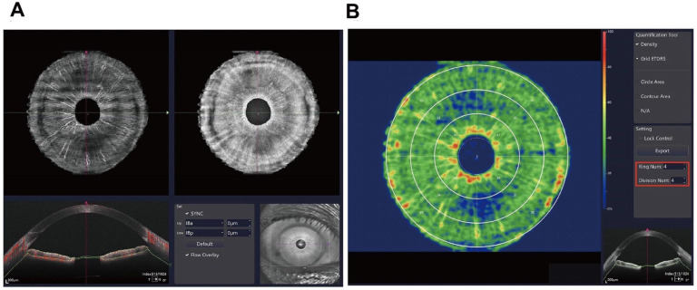
Methods and analysis: In this prospective study, 88 eyes of 44 participants who underwent uneventful cataract surgery were followed up at enrolment, 1 day before, and 1 day, 1 week and 1 month after each eye surgery. Iris blood flow was quantified by a swept-source optical coherence tomography angiography and intraoperative pain was evaluated. Participants were divided into three groups according to time intervals between two eye surgeries: ≥1 to <2 weeks (short interval), ≥2 to ≤4 weeks (medium interval) and greater than 4 weeks (long interval).
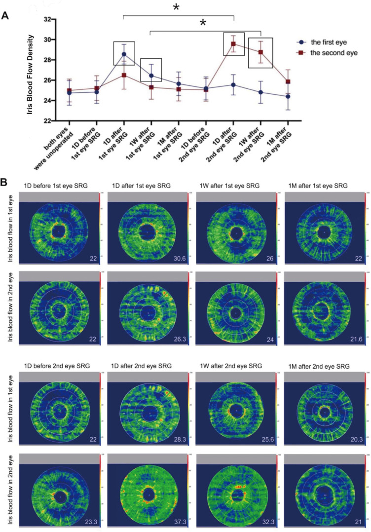
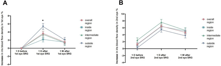
Results: The second eye experienced two significant increases in iris blood flow following cataract surgery: one after the first eye and the other after its own, with the latter being significantly higher than the increase observed in the operated eye after the first surgery (p<0.05). Additionally, the second eye showed higher iris blood flow density in the short interval group compared with the long and medium at specific time points (p<0.05). The bilateral pain index difference positively correlated with second eye iris blood flow density (p<0.05) and was significantly greater in the short interval group than the long interval group (p<0.05).
Conclusions: Iris blood flow increased in the second-operated eye post surgery compared with the first eye, which may correlate to second-eye pain. However, due to the variability in individual pain perception, a larger sample size is needed to prospectively validate our findings to improve their generalisability.

 求助内容:
求助内容: 应助结果提醒方式:


