{"title":"Role of P38 lipoprotein in Mycoplasma penetrans adhesion to human urothelial cells.","authors":"Kaihua Zhang, Weilu Zou, Yanming Chen","doi":"10.1186/s12866-025-04215-w","DOIUrl":null,"url":null,"abstract":"<p><p>Mycoplasma penetrans, a bacterium detected in individuals seropositive for HIV and phylogenetically clustered with M. muris, may contribute to the progression of Acquired Immune Deficiency Syndrome (AIDS). Cellular adhesion is essential for Mycoplasma infection of host cells. M. penetrans exhibits the capacity to adhere to and invade human cells, precipitating diseases of the genital and urinary tracts. However, the proteinaceous mediators of its adhesion remain largely elusive. The P35 family lipoprotein, encoded by the mpl gene, is a prominent surface lipoprotein of M. penetrans. Here, we investigated the role of P38, a member of the P35 family, in the adhesion of M. penetrans to human urothelial cells (SV-HUC-1). We expressed and purified recombinant P38 (rP38) and confirmed its localization using Western blot. Adhesion assays, adhesion inhibition assays, and adhesion competition assays were performed to evaluate the adhesive properties of rP38 and M. penetrans. Our findings indicated that P38 localizes to the cell membrane of M. penetrans. Both rP38 and M. penetrans adhered to SV-HUC-1 cells, with optimal adhesion observed at 60 μg/mL for rP38 and 1 × 10⁷ CCU (Colony-Changing Units)/mL for M. penetrans. Anti-rP38 serum partially inhibited M. penetrans adhesion to SV-HUC-1 cells, and rP38 competed with M. penetrans for binding to SV-HUC-1 cells. These results suggest that P38 may function as an adhesin of M. penetrans, providing insights into its pathogenic mechanisms.</p>","PeriodicalId":9233,"journal":{"name":"BMC Microbiology","volume":"25 1","pages":"566"},"PeriodicalIF":4.2000,"publicationDate":"2025-08-30","publicationTypes":"Journal Article","fieldsOfStudy":null,"isOpenAccess":false,"openAccessPdf":"https://www.ncbi.nlm.nih.gov/pmc/articles/PMC12398969/pdf/","citationCount":"0","resultStr":null,"platform":"Semanticscholar","paperid":null,"PeriodicalName":"BMC Microbiology","FirstCategoryId":"99","ListUrlMain":"https://doi.org/10.1186/s12866-025-04215-w","RegionNum":2,"RegionCategory":"生物学","ArticlePicture":[],"TitleCN":null,"AbstractTextCN":null,"PMCID":null,"EPubDate":"","PubModel":"","JCR":"Q2","JCRName":"MICROBIOLOGY","Score":null,"Total":0}
引用次数: 0
Abstract
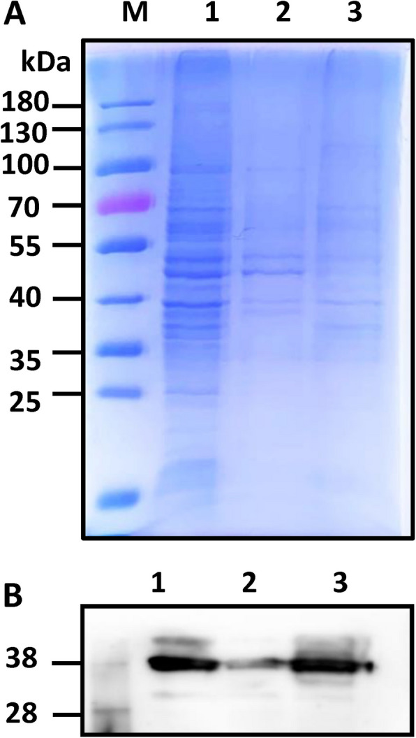
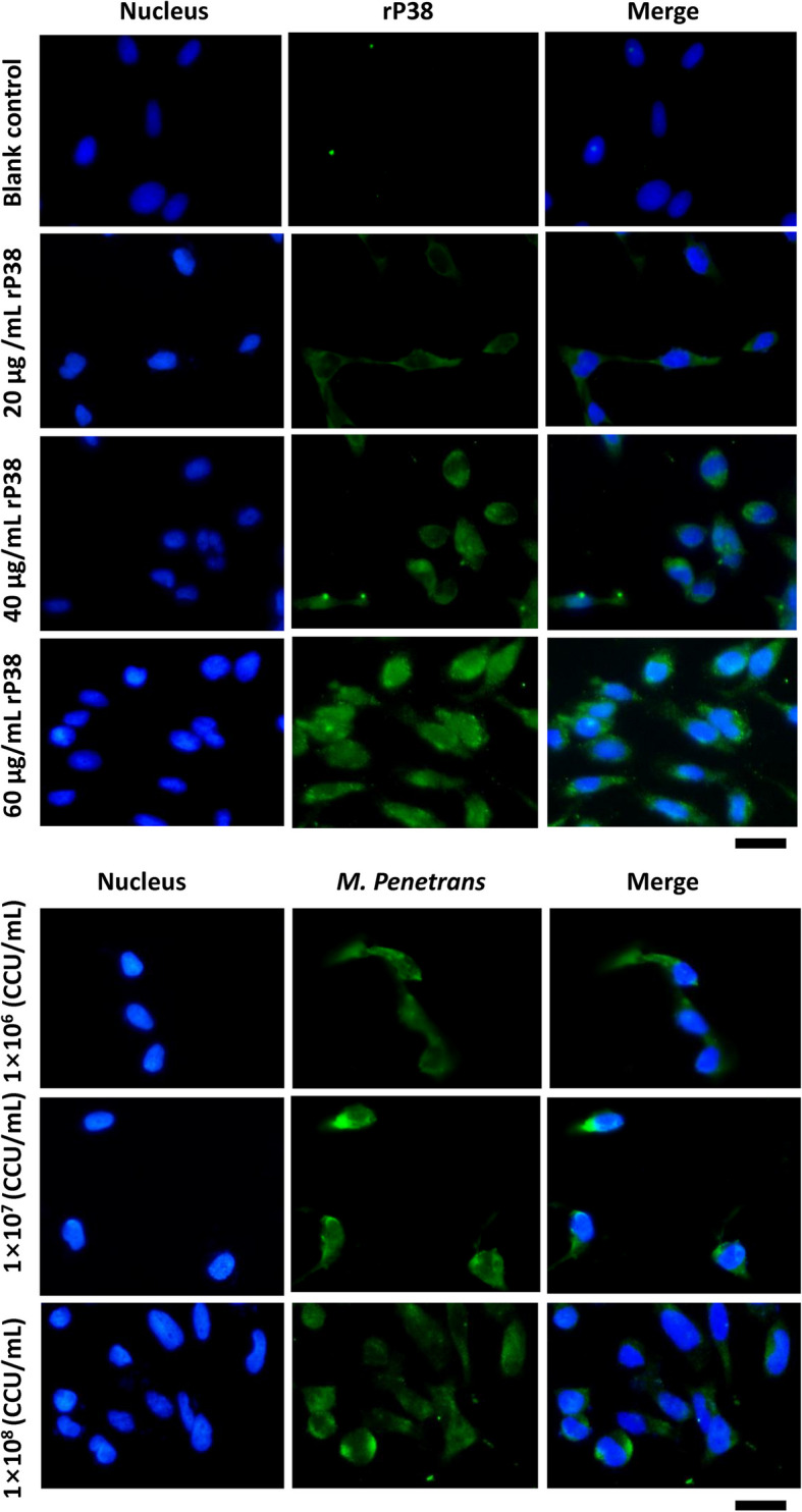
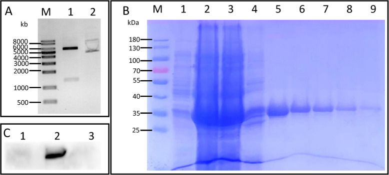
Mycoplasma penetrans, a bacterium detected in individuals seropositive for HIV and phylogenetically clustered with M. muris, may contribute to the progression of Acquired Immune Deficiency Syndrome (AIDS). Cellular adhesion is essential for Mycoplasma infection of host cells. M. penetrans exhibits the capacity to adhere to and invade human cells, precipitating diseases of the genital and urinary tracts. However, the proteinaceous mediators of its adhesion remain largely elusive. The P35 family lipoprotein, encoded by the mpl gene, is a prominent surface lipoprotein of M. penetrans. Here, we investigated the role of P38, a member of the P35 family, in the adhesion of M. penetrans to human urothelial cells (SV-HUC-1). We expressed and purified recombinant P38 (rP38) and confirmed its localization using Western blot. Adhesion assays, adhesion inhibition assays, and adhesion competition assays were performed to evaluate the adhesive properties of rP38 and M. penetrans. Our findings indicated that P38 localizes to the cell membrane of M. penetrans. Both rP38 and M. penetrans adhered to SV-HUC-1 cells, with optimal adhesion observed at 60 μg/mL for rP38 and 1 × 10⁷ CCU (Colony-Changing Units)/mL for M. penetrans. Anti-rP38 serum partially inhibited M. penetrans adhesion to SV-HUC-1 cells, and rP38 competed with M. penetrans for binding to SV-HUC-1 cells. These results suggest that P38 may function as an adhesin of M. penetrans, providing insights into its pathogenic mechanisms.
期刊介绍:
BMC Microbiology is an open access, peer-reviewed journal that considers articles on analytical and functional studies of prokaryotic and eukaryotic microorganisms, viruses and small parasites, as well as host and therapeutic responses to them and their interaction with the environment.
 求助内容:
求助内容: 应助结果提醒方式:


