{"title":"A giant anterior mediastinal thymolipoma mimicking dextrocardia with cardiomegaly: a case report and review of literature.","authors":"Niroshan Lokunarangoda, Dhammike Rasnayake, Miyuru Dulanjana, Duminda Samarasinghe, Ushani Wariyapperuma","doi":"10.1186/s12890-025-03888-3","DOIUrl":null,"url":null,"abstract":"<p><strong>Background: </strong>Thymolipoma is a rare benign anterior mediastinal tumour composed of thymic and adipose tissue, accounting for only 2-9% of thymic neoplasms (Mohamud et al., J Surg Case Rep 2020,2020; Shrivastava and Ntiamoah, Radiol Case Rep 15:1538-1540, 2020). Patients are often asymptomatic, and these masses are often detected incidentally. We present a case of a young male with an anterior mediastinal thymolipoma that initially mimicked dextrocardia and cardiomegaly based on findings from clinical examination and chest radiography.</p><p><strong>Case presentation: </strong>A 22-year-old university student was incidentally noted to have abnormal findings on cardiorespiratory examination. A chest X-ray suggested dextrocardia with cardiomegaly, but further imaging revealed a large anterior mediastinal mass. Contrast-enhanced computed tomography (CT) of the chest showed a well-encapsulated, predominantly fat-density mass (approximately 24 × 15 × 12 cm) in the anterior mediastinum extending into the right hemithorax without invasion of adjacent structures. The lesion caused rightward mediastinal widening but no actual cardiac enlargement, dextroposition or lung collapse. Surgical resection via right mini-thoracotomy was performed. The Gross examination revealed a large, lobulated, encapsulated tumour with a yellow, fatty cut surface and interspersed solid areas. Histopathology confirmed the presence of mature adipose tissue with thymic lobules (cortex, medulla, and Hassall's corpuscles), consistent with thymolipoma, showing no evidence of malignancy. The patient's postoperative recovery was uneventful, and the heart resumed its normal anatomical position.</p><p><strong>Conclusion: </strong>This case highlights that an anterior mediastinal mass, such as a thymolipoma, can masquerade as dextrocardia and cardiomegaly by distorting the mediastinal silhouette. It is essential to distinguish a mediastinal tumour from true congenital dextrocardia or pulmonary conditions, as each requires markedly different management approaches. Thorough imaging and histological evaluation ensured the correct diagnosis and curative surgical treatment. This report emphasises the importance of considering mediastinal pathology in patients with apparent dextrocardia, as detected during examination or imaging.</p>","PeriodicalId":9148,"journal":{"name":"BMC Pulmonary Medicine","volume":"25 1","pages":"418"},"PeriodicalIF":2.8000,"publicationDate":"2025-09-01","publicationTypes":"Journal Article","fieldsOfStudy":null,"isOpenAccess":false,"openAccessPdf":"https://www.ncbi.nlm.nih.gov/pmc/articles/PMC12400621/pdf/","citationCount":"0","resultStr":null,"platform":"Semanticscholar","paperid":null,"PeriodicalName":"BMC Pulmonary Medicine","FirstCategoryId":"3","ListUrlMain":"https://doi.org/10.1186/s12890-025-03888-3","RegionNum":3,"RegionCategory":"医学","ArticlePicture":[],"TitleCN":null,"AbstractTextCN":null,"PMCID":null,"EPubDate":"","PubModel":"","JCR":"Q2","JCRName":"RESPIRATORY SYSTEM","Score":null,"Total":0}
引用次数: 0
Abstract
Background: Thymolipoma is a rare benign anterior mediastinal tumour composed of thymic and adipose tissue, accounting for only 2-9% of thymic neoplasms (Mohamud et al., J Surg Case Rep 2020,2020; Shrivastava and Ntiamoah, Radiol Case Rep 15:1538-1540, 2020). Patients are often asymptomatic, and these masses are often detected incidentally. We present a case of a young male with an anterior mediastinal thymolipoma that initially mimicked dextrocardia and cardiomegaly based on findings from clinical examination and chest radiography.
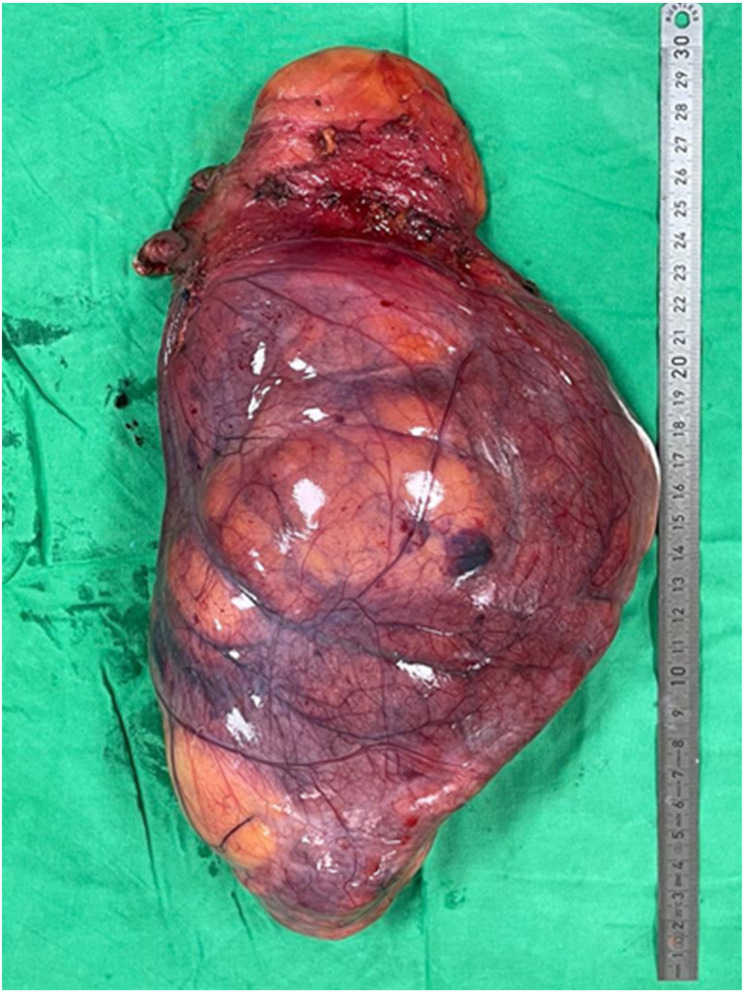
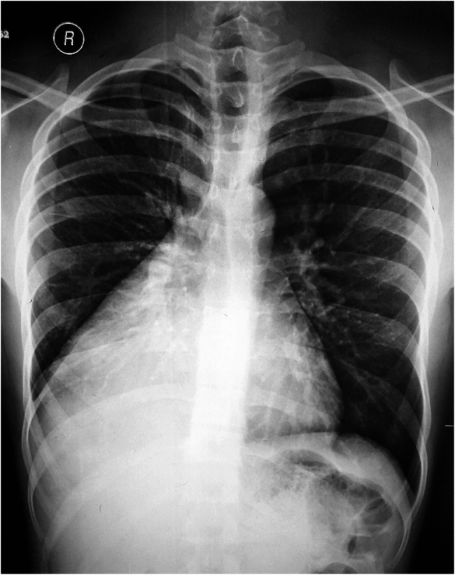
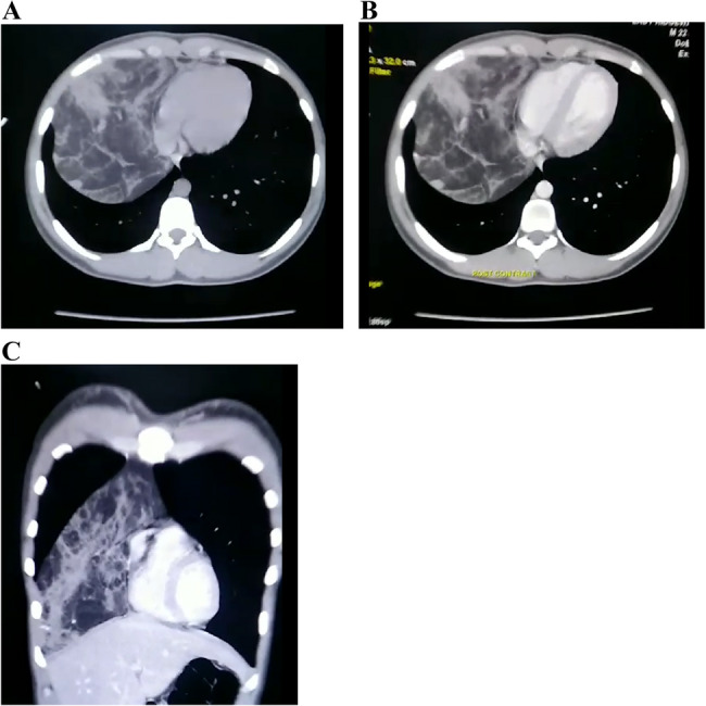
Case presentation: A 22-year-old university student was incidentally noted to have abnormal findings on cardiorespiratory examination. A chest X-ray suggested dextrocardia with cardiomegaly, but further imaging revealed a large anterior mediastinal mass. Contrast-enhanced computed tomography (CT) of the chest showed a well-encapsulated, predominantly fat-density mass (approximately 24 × 15 × 12 cm) in the anterior mediastinum extending into the right hemithorax without invasion of adjacent structures. The lesion caused rightward mediastinal widening but no actual cardiac enlargement, dextroposition or lung collapse. Surgical resection via right mini-thoracotomy was performed. The Gross examination revealed a large, lobulated, encapsulated tumour with a yellow, fatty cut surface and interspersed solid areas. Histopathology confirmed the presence of mature adipose tissue with thymic lobules (cortex, medulla, and Hassall's corpuscles), consistent with thymolipoma, showing no evidence of malignancy. The patient's postoperative recovery was uneventful, and the heart resumed its normal anatomical position.
Conclusion: This case highlights that an anterior mediastinal mass, such as a thymolipoma, can masquerade as dextrocardia and cardiomegaly by distorting the mediastinal silhouette. It is essential to distinguish a mediastinal tumour from true congenital dextrocardia or pulmonary conditions, as each requires markedly different management approaches. Thorough imaging and histological evaluation ensured the correct diagnosis and curative surgical treatment. This report emphasises the importance of considering mediastinal pathology in patients with apparent dextrocardia, as detected during examination or imaging.
背景:胸腺脂肪瘤是一种罕见的由胸腺和脂肪组织组成的前纵隔良性肿瘤,仅占胸腺肿瘤的2-9% (Mohamud et al., J surgery Case Rep 2020,2020; Shrivastava and Ntiamoah, Radiol Case Rep 15:1538-1540, 2020)。患者通常无症状,这些肿块通常是偶然发现的。我们报告一个年轻男性前纵隔胸腺脂肪瘤的病例,根据临床检查和胸部x线检查的结果,最初表现为右心和心脏肥大。病例介绍:一名22岁的大学生在心肺检查中偶然发现异常。胸部x线显示右心伴心脏肿大,但进一步影像学显示前纵隔肿块较大。胸部增强计算机断层扫描(CT)显示前纵隔包裹良好,主要是脂肪密度肿块(约24 × 15 × 12 cm),延伸至右半胸,未侵犯邻近结构。病变引起右侧纵隔增宽,但没有实际的心脏增大、右移或肺萎陷。经右小开胸手术切除。大体检查显示一个大的,分叶的,包裹的肿瘤,有黄色的脂肪切面和散布的实性区域。组织病理学证实成熟脂肪组织与胸腺小叶(皮质、髓质和Hassall小体)的存在,与胸腺脂肪瘤一致,未显示恶性肿瘤的证据。患者术后恢复顺利,心脏恢复正常解剖位置。结论:本病例强调了前纵隔肿块,如胸腺脂肪瘤,可以通过扭曲纵隔轮廓来伪装为右心和心脏肥大。区分纵隔肿瘤与真正的先天性右心或肺部疾病是必要的,因为它们需要明显不同的治疗方法。充分的影像学和组织学检查确保了正确的诊断和有效的手术治疗。本报告强调了在检查或影像学检查中发现的明显右心患者考虑纵隔病理的重要性。
期刊介绍:
BMC Pulmonary Medicine is an open access, peer-reviewed journal that considers articles on all aspects of the prevention, diagnosis and management of pulmonary and associated disorders, as well as related molecular genetics, pathophysiology, and epidemiology.
 求助内容:
求助内容: 应助结果提醒方式:


