{"title":"Intraoperative UBM-guided direct cyclopexy for traumatic cyclodialysis cleft: a four-case series.","authors":"Fei Wang, Xin Jin, Xiang Chen, Bing Chen, Qing-Li Kong, Bao-Ke Hou","doi":"10.1186/s12886-025-04250-1","DOIUrl":null,"url":null,"abstract":"<p><strong>Purpose: </strong>To evaluate the feasibility and effectiveness of intraoperative ultrasonic biomicroscopy (UBM)-guided direct cyclopexy for treating traumatic cyclodialysis clefts.</p><p><strong>Methods: </strong>Intraoperative UBM-guided direct cyclopexy was performed on a total of four eyes. Prior to the conjunctival incision, UBM evaluation was facilitated with a self-designed eye cup. Scleral flaps were generated according to their respective locations. Before the scleral flaps and conjunctiva were closed, UBM was used to promptly assess the repositioning status.</p><p><strong>Results: </strong>1.UBM-guided direct cyclopexies were performed on four male patients (mean age: 39.7 years). In all instances, the cyclodialysis cleft resulted from blunt ocular trauma, with an average time interval between injury and surgical treatment of 29.5 days. 2. A simple cyclodialysis cleft was observed in two patients. One patient had retinal detachment and subluxation of the lens, and one presented with a shallow anterior chamber, iridodialysis, cataract, vitreous hemorrhage, papilledema, and hypotony maculopathy. 3. Prior to surgery, the mean IOP in all four patients was 7 mmHg (range: 6-9 mmHg), which increased to an average of 16.5 mmHg on postsurgical day three (range: 11-22 mmHg). 4. UBM examination confirmed successful closure of the cleft in all patients.</p><p><strong>Conclusion: </strong>During the procedure, UBM-guided direct cyclopexies can accurately assess cyclodialysis cleft repair without being affected by the optical clarity of the refractive media. Preliminary findings demonstrate the feasibility of this approach.</p>","PeriodicalId":9058,"journal":{"name":"BMC Ophthalmology","volume":"25 1","pages":"479"},"PeriodicalIF":1.7000,"publicationDate":"2025-08-21","publicationTypes":"Journal Article","fieldsOfStudy":null,"isOpenAccess":false,"openAccessPdf":"https://www.ncbi.nlm.nih.gov/pmc/articles/PMC12369265/pdf/","citationCount":"0","resultStr":null,"platform":"Semanticscholar","paperid":null,"PeriodicalName":"BMC Ophthalmology","FirstCategoryId":"3","ListUrlMain":"https://doi.org/10.1186/s12886-025-04250-1","RegionNum":4,"RegionCategory":"医学","ArticlePicture":[],"TitleCN":null,"AbstractTextCN":null,"PMCID":null,"EPubDate":"","PubModel":"","JCR":"Q3","JCRName":"OPHTHALMOLOGY","Score":null,"Total":0}
引用次数: 0
Abstract
Purpose: To evaluate the feasibility and effectiveness of intraoperative ultrasonic biomicroscopy (UBM)-guided direct cyclopexy for treating traumatic cyclodialysis clefts.
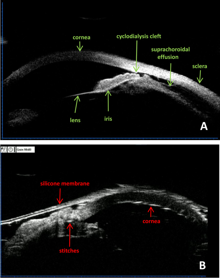
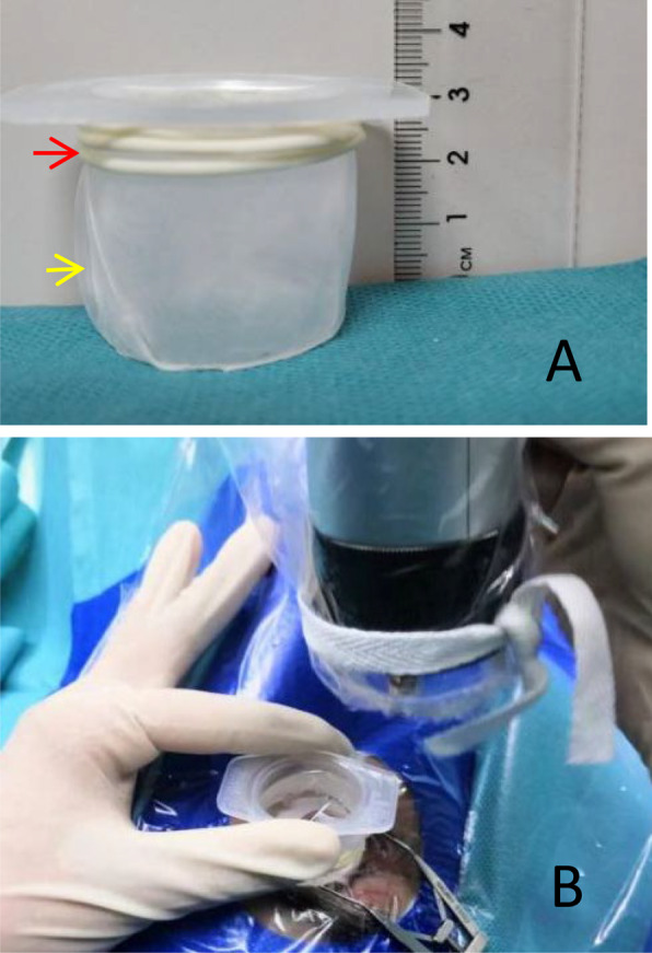
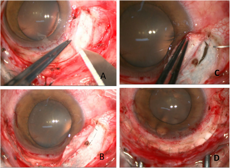
Methods: Intraoperative UBM-guided direct cyclopexy was performed on a total of four eyes. Prior to the conjunctival incision, UBM evaluation was facilitated with a self-designed eye cup. Scleral flaps were generated according to their respective locations. Before the scleral flaps and conjunctiva were closed, UBM was used to promptly assess the repositioning status.
Results: 1.UBM-guided direct cyclopexies were performed on four male patients (mean age: 39.7 years). In all instances, the cyclodialysis cleft resulted from blunt ocular trauma, with an average time interval between injury and surgical treatment of 29.5 days. 2. A simple cyclodialysis cleft was observed in two patients. One patient had retinal detachment and subluxation of the lens, and one presented with a shallow anterior chamber, iridodialysis, cataract, vitreous hemorrhage, papilledema, and hypotony maculopathy. 3. Prior to surgery, the mean IOP in all four patients was 7 mmHg (range: 6-9 mmHg), which increased to an average of 16.5 mmHg on postsurgical day three (range: 11-22 mmHg). 4. UBM examination confirmed successful closure of the cleft in all patients.
Conclusion: During the procedure, UBM-guided direct cyclopexies can accurately assess cyclodialysis cleft repair without being affected by the optical clarity of the refractive media. Preliminary findings demonstrate the feasibility of this approach.
期刊介绍:
BMC Ophthalmology is an open access, peer-reviewed journal that considers articles on all aspects of the prevention, diagnosis and management of eye disorders, as well as related molecular genetics, pathophysiology, and epidemiology.

 求助内容:
求助内容: 应助结果提醒方式:


