{"title":"Ganglion cell layer changes following the idiopathic macular hole surgery using inverted limiting membrane flap technique.","authors":"Mehmet Önen, Muzaffer Şahin, Gökhan Çelik","doi":"10.1186/s12886-025-04317-z","DOIUrl":null,"url":null,"abstract":"<p><strong>Purpose: </strong>The purpose of this study is to compare the ganglion cell layer changes following temporal inverted internal limiting membrane flap (i-ILMF) surgery for idiopathic macular hole (IMH).</p><p><strong>Methods: </strong>This retrospective study included 50 eyes that underwent vitrectomy with a 2.5-disc-diameter temporal inverted internal limiting membrane flap (i-ILMF) technique. Demographic, functional, and anatomical data were collected before and after the surgery. The best corrected visual acuity (BCVA) and optical coherence tomography (OCT) findings such as ganglion cell layer -inner plexiform layer (GCL-IPL) thickness and hole related parameters/indexes were compared in the preoperative period and 6th month after surgery.</p><p><strong>Results: </strong>The average age of the patients was 68.8 ± 10.31 years, and the average duration of visual loss was 10.95 ± 6.54 months. The average GCL-IPL thickness increased significantly from 57.98 ± 21.43 μm to 68.74 ± 13.62 μm at 6 months after surgery (p < 0.001). The nasal GCL-IPL thickness was significantly increased from 56.94 ± 24.18 μm to 73.10 ± 15.39 μm after 6 months after surgery (p < 0.001).</p><p><strong>Conclusion: </strong>The temporal i-ILMF technique not only leads to high anatomical success and visual improvement but also results in a significant increase in GCL-IPL thickness postoperatively, suggesting a unique structural response to this method.</p>","PeriodicalId":9058,"journal":{"name":"BMC Ophthalmology","volume":"25 1","pages":"481"},"PeriodicalIF":1.7000,"publicationDate":"2025-08-21","publicationTypes":"Journal Article","fieldsOfStudy":null,"isOpenAccess":false,"openAccessPdf":"https://www.ncbi.nlm.nih.gov/pmc/articles/PMC12372387/pdf/","citationCount":"0","resultStr":null,"platform":"Semanticscholar","paperid":null,"PeriodicalName":"BMC Ophthalmology","FirstCategoryId":"3","ListUrlMain":"https://doi.org/10.1186/s12886-025-04317-z","RegionNum":4,"RegionCategory":"医学","ArticlePicture":[],"TitleCN":null,"AbstractTextCN":null,"PMCID":null,"EPubDate":"","PubModel":"","JCR":"Q3","JCRName":"OPHTHALMOLOGY","Score":null,"Total":0}
引用次数: 0
Abstract
Purpose: The purpose of this study is to compare the ganglion cell layer changes following temporal inverted internal limiting membrane flap (i-ILMF) surgery for idiopathic macular hole (IMH).
Methods: This retrospective study included 50 eyes that underwent vitrectomy with a 2.5-disc-diameter temporal inverted internal limiting membrane flap (i-ILMF) technique. Demographic, functional, and anatomical data were collected before and after the surgery. The best corrected visual acuity (BCVA) and optical coherence tomography (OCT) findings such as ganglion cell layer -inner plexiform layer (GCL-IPL) thickness and hole related parameters/indexes were compared in the preoperative period and 6th month after surgery.
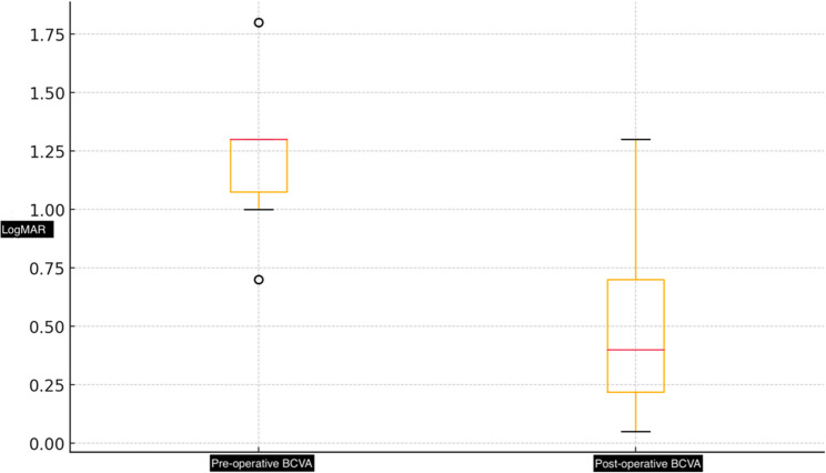
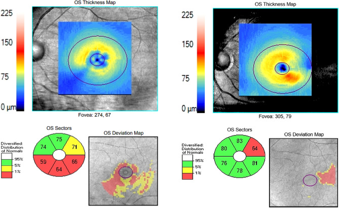
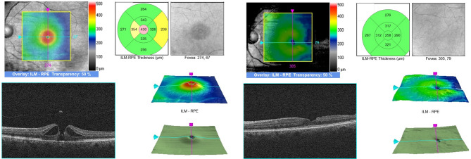
Results: The average age of the patients was 68.8 ± 10.31 years, and the average duration of visual loss was 10.95 ± 6.54 months. The average GCL-IPL thickness increased significantly from 57.98 ± 21.43 μm to 68.74 ± 13.62 μm at 6 months after surgery (p < 0.001). The nasal GCL-IPL thickness was significantly increased from 56.94 ± 24.18 μm to 73.10 ± 15.39 μm after 6 months after surgery (p < 0.001).
Conclusion: The temporal i-ILMF technique not only leads to high anatomical success and visual improvement but also results in a significant increase in GCL-IPL thickness postoperatively, suggesting a unique structural response to this method.
期刊介绍:
BMC Ophthalmology is an open access, peer-reviewed journal that considers articles on all aspects of the prevention, diagnosis and management of eye disorders, as well as related molecular genetics, pathophysiology, and epidemiology.

 求助内容:
求助内容: 应助结果提醒方式:


