Salem Hannoun, Grace Fayad, Nabil K El Ayoubi, Samia J Khoury
{"title":"The effect of lesion filling on brain age estimation in multiple sclerosis.","authors":"Salem Hannoun, Grace Fayad, Nabil K El Ayoubi, Samia J Khoury","doi":"10.1186/s12880-025-01897-6","DOIUrl":null,"url":null,"abstract":"<p><strong>Background: </strong>Brain age estimation is an emerging biomarker for assessing neurodegeneration in multiple sclerosis (MS). However, MS-related lesions can distort structural measurements, potentially leading to inaccuracies in age prediction models. Lesion filling has been proposed as a corrective step, but its impact on brain age estimation and its associations with clinical and structural markers remains unclear.</p><p><strong>Methods: </strong>We analyzed 571 relapsing-remitting MS patients using the BrainAgeR pipeline to estimate brain age from both non-lesion-filled and lesion-filled T1-weighted images. Bias correction was applied to remove age-related prediction bias. Brain Age Gap (BAG) was computed as the difference between corrected predicted brain age and chronological age. Multivariable linear regression models were used to assess associations between BAG and clinical outcomes (EDSS, 9HPT, SDMT, 25FWT) and volumetric measures.</p><p><strong>Results: </strong>Non-lesion-filled and lesion-filled brain age estimates showed excellent agreement (r = 0.97; ICC = 0.962), with a mean difference of 1.23 years and slightly lower mean absolute error for lesion-filled predictions (8.12 vs. 9.40 years). Both BAG measures were significantly associated with EDSS, 9HPT, and SDMT, though effect sizes were modest. Lesion-filled BAG showed stronger and more consistent associations with gray matter, thalamic, and hippocampal volumes, and these associations remained significant after Bonferroni correction.</p><p><strong>Conclusion: </strong>Lesion filling modestly improves structural interpretability of brain age estimates in MS but has limited effect on clinical correlations. The high concordance between lesion-filled and non-lesion-filled estimates confirms the robustness of brain age as a biomarker, while supporting the use of lesion correction when structural precision is essential.</p>","PeriodicalId":9020,"journal":{"name":"BMC Medical Imaging","volume":"25 1","pages":"356"},"PeriodicalIF":3.2000,"publicationDate":"2025-08-27","publicationTypes":"Journal Article","fieldsOfStudy":null,"isOpenAccess":false,"openAccessPdf":"https://www.ncbi.nlm.nih.gov/pmc/articles/PMC12382106/pdf/","citationCount":"0","resultStr":null,"platform":"Semanticscholar","paperid":null,"PeriodicalName":"BMC Medical Imaging","FirstCategoryId":"3","ListUrlMain":"https://doi.org/10.1186/s12880-025-01897-6","RegionNum":3,"RegionCategory":"医学","ArticlePicture":[],"TitleCN":null,"AbstractTextCN":null,"PMCID":null,"EPubDate":"","PubModel":"","JCR":"Q2","JCRName":"RADIOLOGY, NUCLEAR MEDICINE & MEDICAL IMAGING","Score":null,"Total":0}
引用次数: 0
Abstract
Background: Brain age estimation is an emerging biomarker for assessing neurodegeneration in multiple sclerosis (MS). However, MS-related lesions can distort structural measurements, potentially leading to inaccuracies in age prediction models. Lesion filling has been proposed as a corrective step, but its impact on brain age estimation and its associations with clinical and structural markers remains unclear.
Methods: We analyzed 571 relapsing-remitting MS patients using the BrainAgeR pipeline to estimate brain age from both non-lesion-filled and lesion-filled T1-weighted images. Bias correction was applied to remove age-related prediction bias. Brain Age Gap (BAG) was computed as the difference between corrected predicted brain age and chronological age. Multivariable linear regression models were used to assess associations between BAG and clinical outcomes (EDSS, 9HPT, SDMT, 25FWT) and volumetric measures.
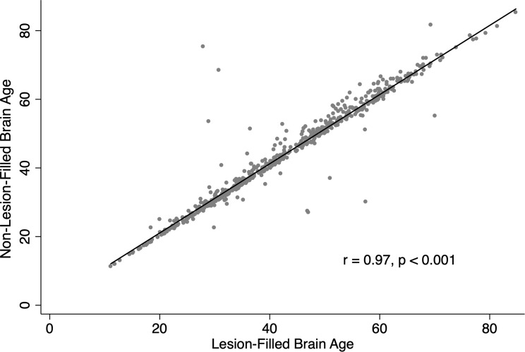
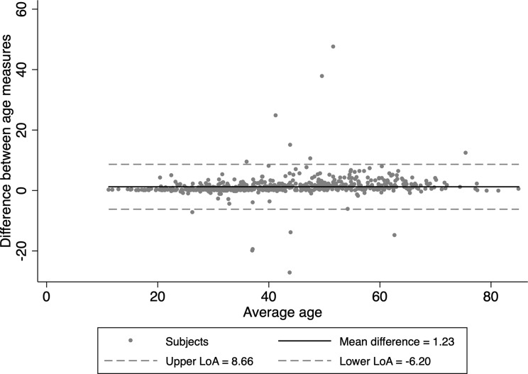
Results: Non-lesion-filled and lesion-filled brain age estimates showed excellent agreement (r = 0.97; ICC = 0.962), with a mean difference of 1.23 years and slightly lower mean absolute error for lesion-filled predictions (8.12 vs. 9.40 years). Both BAG measures were significantly associated with EDSS, 9HPT, and SDMT, though effect sizes were modest. Lesion-filled BAG showed stronger and more consistent associations with gray matter, thalamic, and hippocampal volumes, and these associations remained significant after Bonferroni correction.
Conclusion: Lesion filling modestly improves structural interpretability of brain age estimates in MS but has limited effect on clinical correlations. The high concordance between lesion-filled and non-lesion-filled estimates confirms the robustness of brain age as a biomarker, while supporting the use of lesion correction when structural precision is essential.
期刊介绍:
BMC Medical Imaging is an open access journal publishing original peer-reviewed research articles in the development, evaluation, and use of imaging techniques and image processing tools to diagnose and manage disease.

 求助内容:
求助内容: 应助结果提醒方式:


