Multiparametric MRI for differential diagnosis of primary central nervous system lymphoma and atypical glioblastoma: an analysis incorporating DWI, DCE-MRI, and contrast agent preload DSC-PWI.
IF 3.2 3区 医学Q2 RADIOLOGY, NUCLEAR MEDICINE & MEDICAL IMAGING
{"title":"Multiparametric MRI for differential diagnosis of primary central nervous system lymphoma and atypical glioblastoma: an analysis incorporating DWI, DCE-MRI, and contrast agent preload DSC-PWI.","authors":"Lan Yu, Shujie Yu, Feng Wang, Xiaofang Zhou, Feiman Yang, Dairong Cao, Zhen Xing","doi":"10.1186/s12880-025-01886-9","DOIUrl":null,"url":null,"abstract":"<p><strong>Purpose: </strong>The differential diagnosis of primary central nervous system lymphoma (PCNSL) and atypical glioblastoma (aGBM) exhibiting homogeneous enhancement and negligible necrosis poses a significant challenge for conventional MRI. The study aims to investigate diffusion-weighted imaging (DWI), dynamic contrast-enhanced (DCE) MRI, and contrast agent (CA) preload dynamic susceptibility contrast perfusion-weighted imaging (DSC-PWI) to differentiate aGBM and PCNSL.</p><p><strong>Materials and methods: </strong>This retrospective study analyzed 27 patients with aGBM (solid enhancement without visible necrosis) and 105 patients with PCNSL, all undergoing preoperative DWI, DCE-MRI, and CA preload DSC-PWI. The relative apparent diffusion coefficient (rADC) and relative cerebral blood volume (rCBV) were obtained from DWI and DSC-PWI. The pharmacokinetic parameters (Ktrans, Ve, Kep, and iAUC) were acquired using DCE-MRI. The independent-samples t-test and Mann-Whitney U test were utilized to compare parameters. A binary logistic regression analysis was performed to assess the combined effect of various parameters. Before regression analysis, collinearity analysis of parameters was performed. The diagnostic capability of each parameter and their combination were evaluated by receiver operating characteristic (ROC) with area under the curve (AUC) and compared with DeLong test.</p><p><strong>Results: </strong>In comparison to aGBM, the Ktrans, Ve, and iAUC were significantly elevated in PCNSL, whereas the rCBV and rADC were significantly lower (p < 0.05 for all comparisons). Meanwhile, these parameters allowed excellent diagnostic performance (AUC = 0.817 [rCBV], 0.751 [rADC], 0.808 [Ktrans], 0.765 [Ve], and 0.801 [iAUC]; DeLong test, p > 0.05 for all comparisons). Notably, the combination of all these parameters significantly increased the probability of distinguishing aGBM from PCNSL (AUC = 0.966).</p><p><strong>Conclusions: </strong>DWI, DCE-MRI, and CA preload DSC-PWI can effectively differentiate aGBM from PCNSL, and the combination of all three techniques significantly enhances the discriminatory efficacy.</p>","PeriodicalId":9020,"journal":{"name":"BMC Medical Imaging","volume":"25 1","pages":"345"},"PeriodicalIF":3.2000,"publicationDate":"2025-08-25","publicationTypes":"Journal Article","fieldsOfStudy":null,"isOpenAccess":false,"openAccessPdf":"https://www.ncbi.nlm.nih.gov/pmc/articles/PMC12376417/pdf/","citationCount":"0","resultStr":null,"platform":"Semanticscholar","paperid":null,"PeriodicalName":"BMC Medical Imaging","FirstCategoryId":"3","ListUrlMain":"https://doi.org/10.1186/s12880-025-01886-9","RegionNum":3,"RegionCategory":"医学","ArticlePicture":[],"TitleCN":null,"AbstractTextCN":null,"PMCID":null,"EPubDate":"","PubModel":"","JCR":"Q2","JCRName":"RADIOLOGY, NUCLEAR MEDICINE & MEDICAL IMAGING","Score":null,"Total":0}
引用次数: 0
Abstract
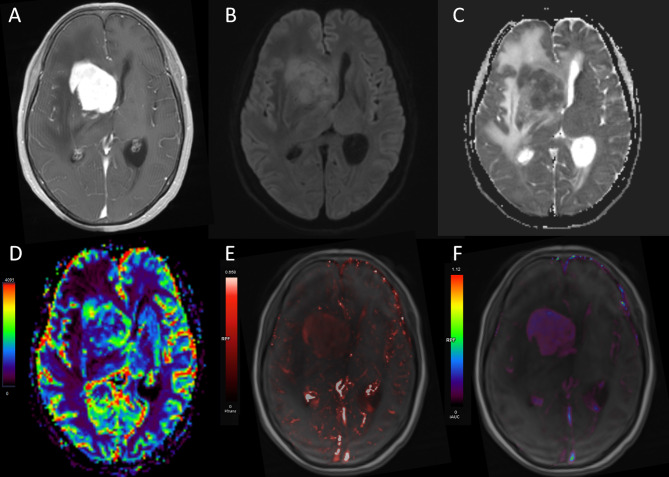
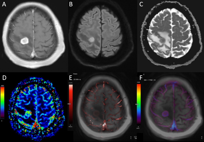
Purpose: The differential diagnosis of primary central nervous system lymphoma (PCNSL) and atypical glioblastoma (aGBM) exhibiting homogeneous enhancement and negligible necrosis poses a significant challenge for conventional MRI. The study aims to investigate diffusion-weighted imaging (DWI), dynamic contrast-enhanced (DCE) MRI, and contrast agent (CA) preload dynamic susceptibility contrast perfusion-weighted imaging (DSC-PWI) to differentiate aGBM and PCNSL.
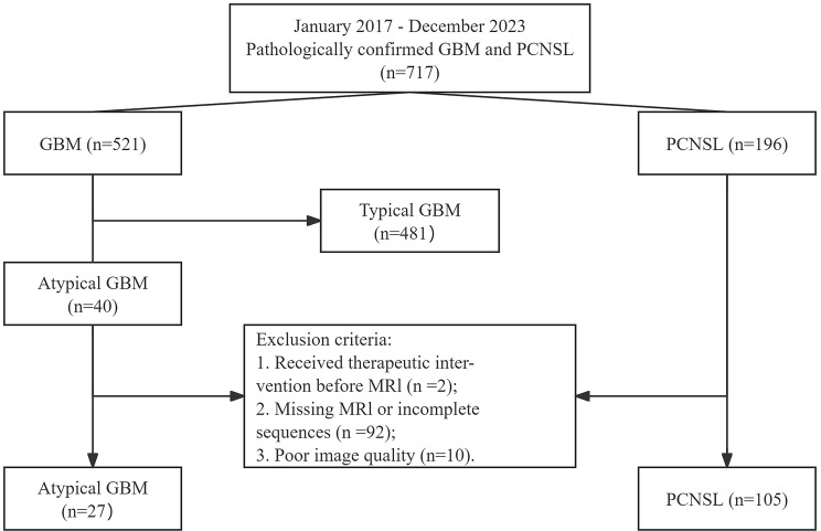
Materials and methods: This retrospective study analyzed 27 patients with aGBM (solid enhancement without visible necrosis) and 105 patients with PCNSL, all undergoing preoperative DWI, DCE-MRI, and CA preload DSC-PWI. The relative apparent diffusion coefficient (rADC) and relative cerebral blood volume (rCBV) were obtained from DWI and DSC-PWI. The pharmacokinetic parameters (Ktrans, Ve, Kep, and iAUC) were acquired using DCE-MRI. The independent-samples t-test and Mann-Whitney U test were utilized to compare parameters. A binary logistic regression analysis was performed to assess the combined effect of various parameters. Before regression analysis, collinearity analysis of parameters was performed. The diagnostic capability of each parameter and their combination were evaluated by receiver operating characteristic (ROC) with area under the curve (AUC) and compared with DeLong test.
Results: In comparison to aGBM, the Ktrans, Ve, and iAUC were significantly elevated in PCNSL, whereas the rCBV and rADC were significantly lower (p < 0.05 for all comparisons). Meanwhile, these parameters allowed excellent diagnostic performance (AUC = 0.817 [rCBV], 0.751 [rADC], 0.808 [Ktrans], 0.765 [Ve], and 0.801 [iAUC]; DeLong test, p > 0.05 for all comparisons). Notably, the combination of all these parameters significantly increased the probability of distinguishing aGBM from PCNSL (AUC = 0.966).
Conclusions: DWI, DCE-MRI, and CA preload DSC-PWI can effectively differentiate aGBM from PCNSL, and the combination of all three techniques significantly enhances the discriminatory efficacy.
期刊介绍:
BMC Medical Imaging is an open access journal publishing original peer-reviewed research articles in the development, evaluation, and use of imaging techniques and image processing tools to diagnose and manage disease.

 求助内容:
求助内容: 应助结果提醒方式:


