Regenerative potential of the intermediate filaments of albino rat parotid glands subjected to fractionated radiotherapy: an immunohistochemical analysis.
Sherif S Hassan, Ehab T Azab, Alaa W Alqutub, Mashael S Alqahtani, Abrar K Demyati, Abdullah A Holdar, Fatma M Alkassimi, Mahmoud A Attia, Reda A Nofal
{"title":"Regenerative potential of the intermediate filaments of albino rat parotid glands subjected to fractionated radiotherapy: an immunohistochemical analysis.","authors":"Sherif S Hassan, Ehab T Azab, Alaa W Alqutub, Mashael S Alqahtani, Abrar K Demyati, Abdullah A Holdar, Fatma M Alkassimi, Mahmoud A Attia, Reda A Nofal","doi":"10.2340/aos.v84.44182","DOIUrl":null,"url":null,"abstract":"<p><strong>Objectives: </strong>Radiotherapy is a common treatment for head and neck malignancies; however, it frequently affects salivary glands, leading to xerostomia. This study evaluated the effects of radiotherapy on cytokeratin localization in the parotid gland, examining whether changes indicate recovery or progressive damage over a year.</p><p><strong>Methods: </strong>The study included eight control rats and 16 irradiated rats exposed to 30 Gy of radiation over 6 days. The experiment was conducted from January 2023 to April 2024. Subgroup IIa rats were sacrificed 1 month after radiation exposure, while subgroup IIb rats were sacrificed after 1 year. The parotid gland was prepared for histological and immunohistochemical analysis of intermediate filaments.</p><p><strong>Results: </strong>In the control parotid gland, immunohistochemical analysis revealed mild cytokeratin in ductal and serous cells. Subgroup IIa exhibited strong cytokeratin expression in the acini and duct cells, which was significantly different from the control group. One year after radiation, the cytokeratin of subgroup IIb was comparable to that of the control, with no significant difference.</p><p><strong>Conclusion: </strong>In subgroup IIa, cytokeratin staining was notably stronger in ductal and acinar cells, leading to disrupted distribution that impaired saliva production and transport. In subgroup IIb, the redistribution of cytokeratin exhibited distinct recovery patterns in ductal and acinar cells.</p>","PeriodicalId":7313,"journal":{"name":"Acta Odontologica Scandinavica","volume":"84 ","pages":"472-478"},"PeriodicalIF":1.9000,"publicationDate":"2025-08-21","publicationTypes":"Journal Article","fieldsOfStudy":null,"isOpenAccess":false,"openAccessPdf":"https://www.ncbi.nlm.nih.gov/pmc/articles/PMC12382380/pdf/","citationCount":"0","resultStr":null,"platform":"Semanticscholar","paperid":null,"PeriodicalName":"Acta Odontologica Scandinavica","FirstCategoryId":"3","ListUrlMain":"https://doi.org/10.2340/aos.v84.44182","RegionNum":4,"RegionCategory":"医学","ArticlePicture":[],"TitleCN":null,"AbstractTextCN":null,"PMCID":null,"EPubDate":"","PubModel":"","JCR":"Q3","JCRName":"DENTISTRY, ORAL SURGERY & MEDICINE","Score":null,"Total":0}
引用次数: 0
Abstract
Objectives: Radiotherapy is a common treatment for head and neck malignancies; however, it frequently affects salivary glands, leading to xerostomia. This study evaluated the effects of radiotherapy on cytokeratin localization in the parotid gland, examining whether changes indicate recovery or progressive damage over a year.
Methods: The study included eight control rats and 16 irradiated rats exposed to 30 Gy of radiation over 6 days. The experiment was conducted from January 2023 to April 2024. Subgroup IIa rats were sacrificed 1 month after radiation exposure, while subgroup IIb rats were sacrificed after 1 year. The parotid gland was prepared for histological and immunohistochemical analysis of intermediate filaments.
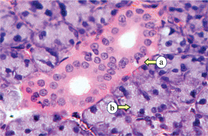
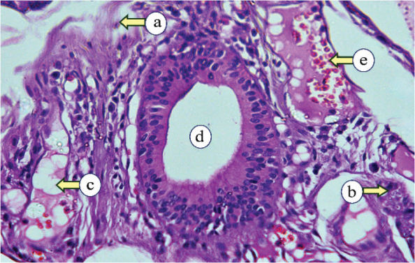
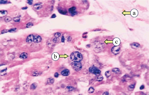
Results: In the control parotid gland, immunohistochemical analysis revealed mild cytokeratin in ductal and serous cells. Subgroup IIa exhibited strong cytokeratin expression in the acini and duct cells, which was significantly different from the control group. One year after radiation, the cytokeratin of subgroup IIb was comparable to that of the control, with no significant difference.
Conclusion: In subgroup IIa, cytokeratin staining was notably stronger in ductal and acinar cells, leading to disrupted distribution that impaired saliva production and transport. In subgroup IIb, the redistribution of cytokeratin exhibited distinct recovery patterns in ductal and acinar cells.
 求助内容:
求助内容: 应助结果提醒方式:


