Jael Soares Batista, Radan Elvis Matias de Oliveira, Wanderson Lucas Alves Dos Santos, Ana Caroline Freitas Caetano de Sousa, Igor Renno Guimarães Lopes, João Augusto Rodrigues Alves Diniz, Thalita Evani Silva de Oliveira, Robério Gomes Olinda, Erick Platini Ferreira de Souto, Moacir Franco de Oliveira
{"title":"An intestinal histiocytic sarcoma in a collared peccary (Pecari tajacu): a case report.","authors":"Jael Soares Batista, Radan Elvis Matias de Oliveira, Wanderson Lucas Alves Dos Santos, Ana Caroline Freitas Caetano de Sousa, Igor Renno Guimarães Lopes, João Augusto Rodrigues Alves Diniz, Thalita Evani Silva de Oliveira, Robério Gomes Olinda, Erick Platini Ferreira de Souto, Moacir Franco de Oliveira","doi":"10.1186/s13028-025-00828-3","DOIUrl":null,"url":null,"abstract":"<p><strong>Background: </strong>Research on cancer in wild animals provides important insights into the mechanisms of carcinogenesis. Histiocytic sarcomas comprise a rare malignant macrophage-dendritic cell lineage neoplasm in wildlife. This study reports a case of histiocytic sarcoma in the small intestine of a collared peccary (Pecari tajacu), describing its clinical, anatomopathological, and immunohistochemical aspects.</p><p><strong>Case presentation: </strong>A six-year-old male collared peccary maintained in captivity at a facility in Northeastern Brazil presented progressive weight loss, diarrhea, anorexia, dyspnea, lethargy, abdominal distension, bristled fur, and pale mucous membranes. A complete blood count indicated a mild degree of anemia and moderate leukocytosis. Treatment included anti-inflammatories and antibiotics; however, on the 18th day after initial presentation, the animal was found dead in its enclosure. An anatomopathological examination revealed that the animal exhibited poor body condition, scant body fat with a gelatinous appearance, hydrothorax, pulmonary edema, and ascites. Thickening of the duodenal wall was observed, along with the presence of a yellowish-white tumor. Histopathological examination of the affected intestinal segment revealed a neoplastic proliferation of round cells with large, hyperchromatic nuclei, prominent nucleoli, and a high mitotic index (20 mitoses per high-power field). Numerous multinucleated and binucleated giant cells were present. The neoplastic cells extensively infiltrated all layers of the intestinal wall, from the mucosa to the serosa. Immunohistochemical analysis showed strong positivity for macrophage/mononuclear phagocytic lineage markers (CD18, IBA-1, and lysozyme), while negative for T-cell (CD3), B-cell (CD79), and plasma cell (MUM1) markers. The proliferation index assessed by Ki-67 was approximately 60%.</p><p><strong>Conclusions: </strong>The histopathological and immunohistochemical findings confirmed the diagnosis of intestinal histiocytic sarcoma in a collared peccary, representing the first documented case of this neoplasm in this species.</p>","PeriodicalId":7181,"journal":{"name":"Acta Veterinaria Scandinavica","volume":"67 1","pages":"43"},"PeriodicalIF":1.7000,"publicationDate":"2025-09-02","publicationTypes":"Journal Article","fieldsOfStudy":null,"isOpenAccess":false,"openAccessPdf":"https://www.ncbi.nlm.nih.gov/pmc/articles/PMC12403295/pdf/","citationCount":"0","resultStr":null,"platform":"Semanticscholar","paperid":null,"PeriodicalName":"Acta Veterinaria Scandinavica","FirstCategoryId":"97","ListUrlMain":"https://doi.org/10.1186/s13028-025-00828-3","RegionNum":2,"RegionCategory":"农林科学","ArticlePicture":[],"TitleCN":null,"AbstractTextCN":null,"PMCID":null,"EPubDate":"","PubModel":"","JCR":"Q2","JCRName":"VETERINARY SCIENCES","Score":null,"Total":0}
引用次数: 0
Abstract
Background: Research on cancer in wild animals provides important insights into the mechanisms of carcinogenesis. Histiocytic sarcomas comprise a rare malignant macrophage-dendritic cell lineage neoplasm in wildlife. This study reports a case of histiocytic sarcoma in the small intestine of a collared peccary (Pecari tajacu), describing its clinical, anatomopathological, and immunohistochemical aspects.
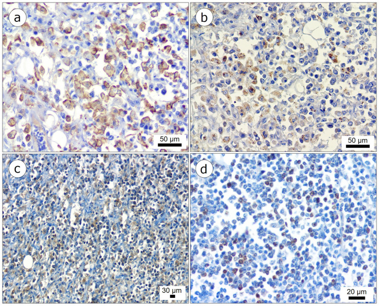
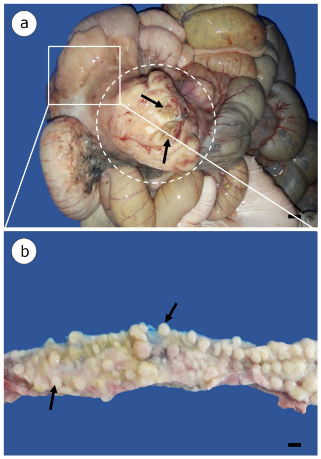
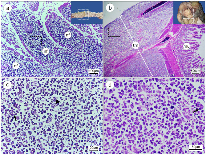
Case presentation: A six-year-old male collared peccary maintained in captivity at a facility in Northeastern Brazil presented progressive weight loss, diarrhea, anorexia, dyspnea, lethargy, abdominal distension, bristled fur, and pale mucous membranes. A complete blood count indicated a mild degree of anemia and moderate leukocytosis. Treatment included anti-inflammatories and antibiotics; however, on the 18th day after initial presentation, the animal was found dead in its enclosure. An anatomopathological examination revealed that the animal exhibited poor body condition, scant body fat with a gelatinous appearance, hydrothorax, pulmonary edema, and ascites. Thickening of the duodenal wall was observed, along with the presence of a yellowish-white tumor. Histopathological examination of the affected intestinal segment revealed a neoplastic proliferation of round cells with large, hyperchromatic nuclei, prominent nucleoli, and a high mitotic index (20 mitoses per high-power field). Numerous multinucleated and binucleated giant cells were present. The neoplastic cells extensively infiltrated all layers of the intestinal wall, from the mucosa to the serosa. Immunohistochemical analysis showed strong positivity for macrophage/mononuclear phagocytic lineage markers (CD18, IBA-1, and lysozyme), while negative for T-cell (CD3), B-cell (CD79), and plasma cell (MUM1) markers. The proliferation index assessed by Ki-67 was approximately 60%.
Conclusions: The histopathological and immunohistochemical findings confirmed the diagnosis of intestinal histiocytic sarcoma in a collared peccary, representing the first documented case of this neoplasm in this species.
期刊介绍:
Acta Veterinaria Scandinavica is an open access journal encompassing all aspects of veterinary research and medicine of domestic and wild animals.

 求助内容:
求助内容: 应助结果提醒方式:


