Jong-Min Jeong, Sung Eun Choi, Young-Ri Shim, Hee-Hoon Kim, Young-Sun Lee, Keungmo Yang, Kyurae Kim, Min Jeong Kim, Katherine Po Sin Chung, Seok-Hwan Kim, Jin-Seok Byun, Hyuk Soo Eun, Won-Il Jeong
{"title":"CX3CR1+ macrophages interact with HSCs to promote HCC through CD8+ T-cell suppression.","authors":"Jong-Min Jeong, Sung Eun Choi, Young-Ri Shim, Hee-Hoon Kim, Young-Sun Lee, Keungmo Yang, Kyurae Kim, Min Jeong Kim, Katherine Po Sin Chung, Seok-Hwan Kim, Jin-Seok Byun, Hyuk Soo Eun, Won-Il Jeong","doi":"10.1097/HEP.0000000000001021","DOIUrl":null,"url":null,"abstract":"<p><strong>Background and aims: </strong>HSCs contribute to HCC progression by regulating multiple factors. However, the entire immunoregulatory functions of HSCs are still obscure. Here, we aim to investigate whether HSCs impose CX3CR1+ macrophages to protumorigenic properties in the peritumoral area.</p><p><strong>Approach and results: </strong>In single-cell RNA-sequencing analysis of patients with HCC, a subpopulation of macrophages specifically expressed Arg1 and Cx3cr1 in the peritumoral area and were highly enriched with retinol metabolism-related genes. Flow cytometry analysis showed significantly increased frequencies of CD14+CD11b+HLA-DR- macrophages with CX3CR1 in the HCC adjacent region where α-smooth muscle actin-expressing activated hepatic stellate cells (aHSCs) showed colocalized expression of CX3CL1. Accordingly, in tumor-bearing mice, Cx3cl1 mRNA expression was notably increased in aHSCs within the adjacent HCC, where infiltration of CX3CR1+Ly6C+ macrophages was mostly observed with decreased CD8+ T cells. In adoptive transfer and in vitro coculture of myeloid cells, we demonstrated that CX3CR1+Ly6C+ macrophages migrated and highly expressed arginase-1 by interacting with retinoid-enriched aHSCs in the adjacent HCC. Direct treatment of retinoids or coculturing with retinol-storing mouse aHSCs or human LX-2 cells significantly increased arginase-1 expression in CX3CR1+Ly6C+ macrophages and human blood CD14+ cells, leading to the suppression of CD8+ T-cell proliferation. Moreover, genetic deficiency of CX3CR1 in myeloid cells or pharmacological inhibition of retinol metabolism remarkably attenuated HCC development.</p><p><strong>Conclusions: </strong>We showed that CX3CR1+Ly6C+ macrophages migrate and interact with aHSCs in the peritumoral region where retinoids induce arginase-1 expression in CX3CR1+Ly6C+ macrophages, subsequently depriving CD8+ T cells of arginine and promoting HCC.</p>","PeriodicalId":177,"journal":{"name":"Hepatology","volume":"82 3","pages":"655-668"},"PeriodicalIF":15.8000,"publicationDate":"2025-09-01","publicationTypes":"Journal Article","fieldsOfStudy":null,"isOpenAccess":false,"openAccessPdf":"https://www.ncbi.nlm.nih.gov/pmc/articles/PMC12356560/pdf/","citationCount":"0","resultStr":null,"platform":"Semanticscholar","paperid":null,"PeriodicalName":"Hepatology","FirstCategoryId":"3","ListUrlMain":"https://doi.org/10.1097/HEP.0000000000001021","RegionNum":1,"RegionCategory":"医学","ArticlePicture":[],"TitleCN":null,"AbstractTextCN":null,"PMCID":null,"EPubDate":"2024/7/19 0:00:00","PubModel":"Epub","JCR":"Q1","JCRName":"GASTROENTEROLOGY & HEPATOLOGY","Score":null,"Total":0}
引用次数: 0
Abstract
Background and aims: HSCs contribute to HCC progression by regulating multiple factors. However, the entire immunoregulatory functions of HSCs are still obscure. Here, we aim to investigate whether HSCs impose CX3CR1+ macrophages to protumorigenic properties in the peritumoral area.
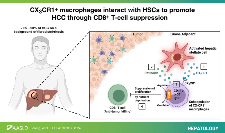
Approach and results: In single-cell RNA-sequencing analysis of patients with HCC, a subpopulation of macrophages specifically expressed Arg1 and Cx3cr1 in the peritumoral area and were highly enriched with retinol metabolism-related genes. Flow cytometry analysis showed significantly increased frequencies of CD14+CD11b+HLA-DR- macrophages with CX3CR1 in the HCC adjacent region where α-smooth muscle actin-expressing activated hepatic stellate cells (aHSCs) showed colocalized expression of CX3CL1. Accordingly, in tumor-bearing mice, Cx3cl1 mRNA expression was notably increased in aHSCs within the adjacent HCC, where infiltration of CX3CR1+Ly6C+ macrophages was mostly observed with decreased CD8+ T cells. In adoptive transfer and in vitro coculture of myeloid cells, we demonstrated that CX3CR1+Ly6C+ macrophages migrated and highly expressed arginase-1 by interacting with retinoid-enriched aHSCs in the adjacent HCC. Direct treatment of retinoids or coculturing with retinol-storing mouse aHSCs or human LX-2 cells significantly increased arginase-1 expression in CX3CR1+Ly6C+ macrophages and human blood CD14+ cells, leading to the suppression of CD8+ T-cell proliferation. Moreover, genetic deficiency of CX3CR1 in myeloid cells or pharmacological inhibition of retinol metabolism remarkably attenuated HCC development.
Conclusions: We showed that CX3CR1+Ly6C+ macrophages migrate and interact with aHSCs in the peritumoral region where retinoids induce arginase-1 expression in CX3CR1+Ly6C+ macrophages, subsequently depriving CD8+ T cells of arginine and promoting HCC.
期刊介绍:
HEPATOLOGY is recognized as the leading publication in the field of liver disease. It features original, peer-reviewed articles covering various aspects of liver structure, function, and disease. The journal's distinguished Editorial Board carefully selects the best articles each month, focusing on topics including immunology, chronic hepatitis, viral hepatitis, cirrhosis, genetic and metabolic liver diseases, liver cancer, and drug metabolism.
 求助内容:
求助内容: 应助结果提醒方式:


