Francesca Borrelli, Giusy Giugliano, Emilie Houliez, Jaromir Behal, Daniele Pirone, Leonilde Roselli, Angela Sardo, Valerio Zupo, Maria Costantini, Lisa Miccio, Pasquale Memmolo, Vittorio Bianco and Pietro Ferraro
{"title":"3D holographic flow cytometry measurements of microalgae: strategies for angle recovery in complex rotation patterns","authors":"Francesca Borrelli, Giusy Giugliano, Emilie Houliez, Jaromir Behal, Daniele Pirone, Leonilde Roselli, Angela Sardo, Valerio Zupo, Maria Costantini, Lisa Miccio, Pasquale Memmolo, Vittorio Bianco and Pietro Ferraro","doi":"10.1039/D5LC00559K","DOIUrl":null,"url":null,"abstract":"<p >Marine ecosystems are in the spotlight, because environmental changes are threatening biodiversity and ecological functions. In this context, microalgae play key ecological roles both in planktonic and benthic ecosystems. Consequently, they are considered indispensable targets for global monitoring programs. However, due to their high spatial and temporal variability and to difficulties of species identification (still relying on microscopy observations), the assessment of roles played by these components of marine ecosystems is demanding. In addition, technologies for a 3D assessment of their complex morphology are scarcely available. Here, we present a comprehensive workflow for retrieving 3D information on microalgae with diverse geometries through holographic microscopy operating in flow-cytometry mode onboard a lab on a chip device. Depending on the rotation patterns of samples, a tailored approach is used to retrieve their rolling angles. We demonstrate the feasibility of measuring 3D data of various microalgae, contingent on the intrinsic optical properties of cells. Specifically, we show that for quasi-transparent and low-scattering microorganisms, the retrieved angles permit quantitative 3D tomographic refractive index (RI) mapping to be achieved, providing full characterization of the alga in terms of its inner structure and outer shape. Moreover, even in the most challenging scenarios, where microalgae exhibit high light absorption or strong scattering, quantitative 3D shape reconstructions of diatoms and dinoflagellates can be at least achieved. Finally, we compare our direct 3D measurements with 2D inferences of 3D properties, obtained using a commercially available microscopy system. The ability to non-invasively obtain 3D information on microalgae marks a fundamental advancement in the field, unlocking a wealth of novel biological insights for characterizing aquatic ecosystems.</p>","PeriodicalId":85,"journal":{"name":"Lab on a Chip","volume":" 20","pages":" 5283-5291"},"PeriodicalIF":5.4000,"publicationDate":"2025-08-18","publicationTypes":"Journal Article","fieldsOfStudy":null,"isOpenAccess":false,"openAccessPdf":"https://pubs.rsc.org/en/content/articlepdf/2025/lc/d5lc00559k?page=search","citationCount":"0","resultStr":null,"platform":"Semanticscholar","paperid":null,"PeriodicalName":"Lab on a Chip","FirstCategoryId":"5","ListUrlMain":"https://pubs.rsc.org/en/content/articlelanding/2025/lc/d5lc00559k","RegionNum":2,"RegionCategory":"工程技术","ArticlePicture":[],"TitleCN":null,"AbstractTextCN":null,"PMCID":null,"EPubDate":"","PubModel":"","JCR":"Q1","JCRName":"BIOCHEMICAL RESEARCH METHODS","Score":null,"Total":0}
引用次数: 0
Abstract
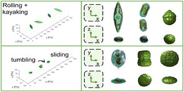
Marine ecosystems are in the spotlight, because environmental changes are threatening biodiversity and ecological functions. In this context, microalgae play key ecological roles both in planktonic and benthic ecosystems. Consequently, they are considered indispensable targets for global monitoring programs. However, due to their high spatial and temporal variability and to difficulties of species identification (still relying on microscopy observations), the assessment of roles played by these components of marine ecosystems is demanding. In addition, technologies for a 3D assessment of their complex morphology are scarcely available. Here, we present a comprehensive workflow for retrieving 3D information on microalgae with diverse geometries through holographic microscopy operating in flow-cytometry mode onboard a lab on a chip device. Depending on the rotation patterns of samples, a tailored approach is used to retrieve their rolling angles. We demonstrate the feasibility of measuring 3D data of various microalgae, contingent on the intrinsic optical properties of cells. Specifically, we show that for quasi-transparent and low-scattering microorganisms, the retrieved angles permit quantitative 3D tomographic refractive index (RI) mapping to be achieved, providing full characterization of the alga in terms of its inner structure and outer shape. Moreover, even in the most challenging scenarios, where microalgae exhibit high light absorption or strong scattering, quantitative 3D shape reconstructions of diatoms and dinoflagellates can be at least achieved. Finally, we compare our direct 3D measurements with 2D inferences of 3D properties, obtained using a commercially available microscopy system. The ability to non-invasively obtain 3D information on microalgae marks a fundamental advancement in the field, unlocking a wealth of novel biological insights for characterizing aquatic ecosystems.
期刊介绍:
Lab on a Chip is the premiere journal that publishes cutting-edge research in the field of miniaturization. By their very nature, microfluidic/nanofluidic/miniaturized systems are at the intersection of disciplines, spanning fundamental research to high-end application, which is reflected by the broad readership of the journal. Lab on a Chip publishes two types of papers on original research: full-length research papers and communications. Papers should demonstrate innovations, which can come from technical advancements or applications addressing pressing needs in globally important areas. The journal also publishes Comments, Reviews, and Perspectives.
 求助内容:
求助内容: 应助结果提醒方式:


