Olga V. Zaitseva, Roman V. Smirnov, Sergey A. Petrov, Anatoly A. Petrov
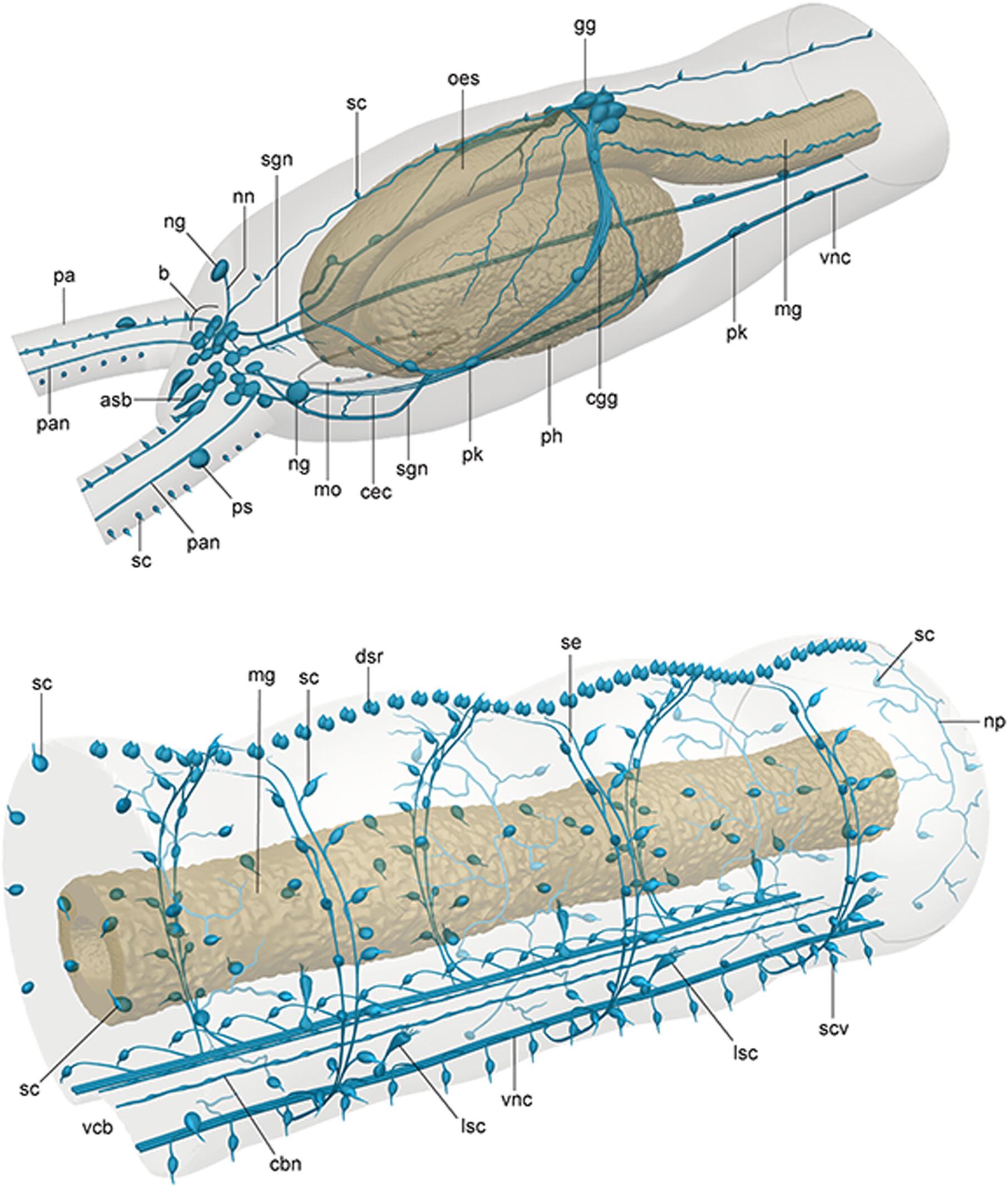
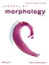
{"title":"Morphological Organization, Sensory Structures and Catecholaminergic Nervous System of Lindrilus flavocapitatus (Annelida: Protodrilidae)","authors":"Olga V. Zaitseva, Roman V. Smirnov, Sergey A. Petrov, Anatoly A. Petrov","doi":"10.1002/jmor.70079","DOIUrl":null,"url":null,"abstract":"<div>\n \n <p>Protodrilidae is a small family of almost exclusively interstitial annelids that lack parapodia and chaetae and possess a basiepithelial nervous system. This study presents a histological description of <i>Lindrilus flavocapitatus</i> (Uljanin, 1877), a protodrilid species last examined morphologically in the early 20th century, and provides detailed information on the organization of its nervous and sensory systems using histochemical detection of catecholamines (CAs), scanning electron microscopy (SEM), and alpha-tubulin immunolabelling. The epidermal ciliary structures on the head show a species-specific distribution pattern, and SEM reveals three types of ciliary sensory structures, similar to those previously described in other protodrilids. Numerous CA-containing (CAc) cells are found in both central (CNS) and peripheral nervous systems. A spatial correlation between epidermal ciliary structures and CAc cells offers the first direct evidence supporting the sensory function of some known ciliary types and allows hypotheses regarding their sensory modalities. The widespread, mostly diffuse distribution of epidermal CAc cells throughout the trunk, pygidium, and palps suggests a mechanosensory function, although some presumed mechanosensory cells are not catecholaminergic or lack CAs. The presence of CAs in putative phaosomes on the palps also points to a possible role for these neurotransmitters in photoreception. In addition to typical annelid sensory organs such as palpal receptors, nuchal organs, and possible phaosomes, <i>L. flavocapitatus</i> possesses a unique bud-shaped sensory organ and a dorsal ridge-like array of receptor cells, both containing CAs. A prominent CAc gastroesophageal ganglion innervating the complex pharyngeal apparatus of <i>L. flavocapitatus</i> is described for the first time in protodrilids. The results reveal a more differentiated neural and sensory organization in protodrilids than previously recognized. Despite its small body size and a relatively low neuron count, <i>L. flavocapitatus</i> possesses additional CNS regions beyond those common to most annelids and a uniquely organized apical sensory organ.</p></div>","PeriodicalId":16528,"journal":{"name":"Journal of Morphology","volume":"286 9","pages":""},"PeriodicalIF":1.4000,"publicationDate":"2025-09-02","publicationTypes":"Journal Article","fieldsOfStudy":null,"isOpenAccess":false,"openAccessPdf":"","citationCount":"0","resultStr":null,"platform":"Semanticscholar","paperid":null,"PeriodicalName":"Journal of Morphology","FirstCategoryId":"3","ListUrlMain":"https://onlinelibrary.wiley.com/doi/10.1002/jmor.70079","RegionNum":4,"RegionCategory":"医学","ArticlePicture":[],"TitleCN":null,"AbstractTextCN":null,"PMCID":null,"EPubDate":"","PubModel":"","JCR":"Q2","JCRName":"ANATOMY & MORPHOLOGY","Score":null,"Total":0}
引用次数: 0
Abstract
Protodrilidae is a small family of almost exclusively interstitial annelids that lack parapodia and chaetae and possess a basiepithelial nervous system. This study presents a histological description of Lindrilus flavocapitatus (Uljanin, 1877), a protodrilid species last examined morphologically in the early 20th century, and provides detailed information on the organization of its nervous and sensory systems using histochemical detection of catecholamines (CAs), scanning electron microscopy (SEM), and alpha-tubulin immunolabelling. The epidermal ciliary structures on the head show a species-specific distribution pattern, and SEM reveals three types of ciliary sensory structures, similar to those previously described in other protodrilids. Numerous CA-containing (CAc) cells are found in both central (CNS) and peripheral nervous systems. A spatial correlation between epidermal ciliary structures and CAc cells offers the first direct evidence supporting the sensory function of some known ciliary types and allows hypotheses regarding their sensory modalities. The widespread, mostly diffuse distribution of epidermal CAc cells throughout the trunk, pygidium, and palps suggests a mechanosensory function, although some presumed mechanosensory cells are not catecholaminergic or lack CAs. The presence of CAs in putative phaosomes on the palps also points to a possible role for these neurotransmitters in photoreception. In addition to typical annelid sensory organs such as palpal receptors, nuchal organs, and possible phaosomes, L. flavocapitatus possesses a unique bud-shaped sensory organ and a dorsal ridge-like array of receptor cells, both containing CAs. A prominent CAc gastroesophageal ganglion innervating the complex pharyngeal apparatus of L. flavocapitatus is described for the first time in protodrilids. The results reveal a more differentiated neural and sensory organization in protodrilids than previously recognized. Despite its small body size and a relatively low neuron count, L. flavocapitatus possesses additional CNS regions beyond those common to most annelids and a uniquely organized apical sensory organ.
期刊介绍:
The Journal of Morphology welcomes articles of original research in cytology, protozoology, embryology, and general morphology. Articles generally should not exceed 35 printed pages. Preliminary notices or articles of a purely descriptive morphological or taxonomic nature are not included. No paper which has already been published will be accepted, nor will simultaneous publications elsewhere be allowed.
The Journal of Morphology publishes research in functional, comparative, evolutionary and developmental morphology from vertebrates and invertebrates. Human and veterinary anatomy or paleontology are considered when an explicit connection to neontological animal morphology is presented, and the paper contains relevant information for the community of animal morphologists. Based on our long tradition, we continue to seek publishing the best papers in animal morphology.
 求助内容:
求助内容: 应助结果提醒方式:


