Distribution and activation of nitrergic neurons in response to aversive stimulus exposure in the Tropidurus hispidus lizard: Involvement of glutamatergic circuitry
IF 3.2
2区 生物学
Q2 BIOCHEMISTRY & MOLECULAR BIOLOGY
引用次数: 0
Abstract
Introduction
Fear is a response to real aversive stimuli. Studies on phylogenetically distant species like reptiles can offer valuable insights into the neural mechanisms of fear.
Objective
To investigate the activation and distribution of nitrergic neurons in the telencephalon of Tropidurus hispidus lizards and evaluate the role of glutamatergic modulation via NMDA receptors following exposure to an aversive stimulus.
Methods
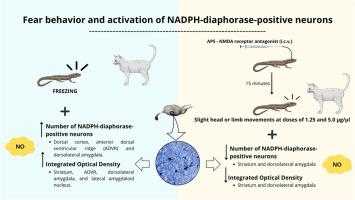
Lizards were exposed to a live cat, and 24 h later, NADPH-diaphorase histochemistry was performed in the telencephalon to quantify neuron number and optical density. In the second stage, animals received i.c.v. injections of the NMDA receptor antagonist AP5 (1.25, 2.5, or 5.0 μg/μl), followed by cat exposure and histochemical analysis.
Results
Exposure to the cat led to increased freezing time in lizards. These animals also showed an increased number of nitrergic neurons in the dorsal cortex, anterior dorsal ventricular ridge (ADVR), and dorsolateral amygdala, as well as elevated integrated optical density (IOD) in the striatum, ADVR, dorsolateral amygdala and lateral amygdaloid nucleus. The AP5 1.25 and 5.0 μg/μl groups exhibited some head or limb movements even in the presence of the cat. The 1.25 μg/μl group showed reduced neuron counts and IOD in the dorsolateral amygdala; the 2.5 μg/μl and 5.0 μg/μl groups showed reduced IOD and neuron counts in the striatum.
Conclusion
Tropidurus hispidus lizards show fear-like behavior and decreased exploration after aversive stimuli, with nitric oxide in the telencephalon – particularly in the striatum and dorsolateral amygdala – modulating this response via NMDA receptor activation.
对厌恶刺激暴露的反应中氮能神经元的分布和激活:谷氨酸能回路的参与
恐惧是对真实的厌恶刺激的反应。对爬行动物等系统发育较远的物种的研究可以为恐惧的神经机制提供有价值的见解。目的观察棘圆蜥远脑氮能神经元的激活和分布,并探讨NMDA受体在不良刺激下对谷氨酸能的调节作用。方法将幼崽暴露于活猫体内,24 h后对远脑进行NADPH-diaphorase组织化学测定神经元数量和光密度。在第二阶段,动物接受静脉注射NMDA受体拮抗剂AP5(1.25、2.5或5.0 μg/μl),然后进行猫暴露和组织化学分析。结果暴露在猫的环境中会增加蜥蜴的冷冻时间。这些动物的背皮质、前背侧脑室脊(ADVR)和杏仁核背外侧的氮能神经元数量增加,纹状体、前背侧脑室脊(ADVR)、杏仁核背外侧和杏仁核外侧的综合光密度(IOD)升高。AP5 1.25和5.0 μl组即使在猫在场的情况下也表现出一定的头部或肢体运动。1.25 μg/μl组杏仁核背外侧神经元数量和IOD减少;2.5 μg/ l和5.0 μg/ l组纹状体的IOD和神经元数量减少。结论棘圆蜥在受到厌恶刺激后表现出类似恐惧的行为,并减少探索活动,这是由于端脑(尤其是纹状体和杏仁核背外侧)的一氧化氮通过NMDA受体激活来调节这种反应。
本文章由计算机程序翻译,如有差异,请以英文原文为准。
求助全文
约1分钟内获得全文
求助全文
来源期刊
Nitric oxide : biology and chemistry
生物-生化与分子生物学
CiteScore
7.50
自引率
7.70%
发文量
74
审稿时长
52 days
期刊介绍:
Nitric Oxide includes original research, methodology papers and reviews relating to nitric oxide and other gasotransmitters such as hydrogen sulfide and carbon monoxide. Special emphasis is placed on the biological chemistry, physiology, pharmacology, enzymology and pathological significance of these molecules in human health and disease. The journal also accepts manuscripts relating to plant and microbial studies involving these molecules.
 求助内容:
求助内容: 应助结果提醒方式:


