Assessing the utility of SISCOM and PISCOM in mapping the epileptogenic zone for epilepsy surgery: A comparative outcome-based study
IF 1.8
4区 医学
Q3 CLINICAL NEUROLOGY
引用次数: 0
Abstract
Intro
Ictal SPECT imaging stands out among non-invasive tools as the primary presurgical method for spatially defining the seizure network. Advanced image analysis methods like SISCOM and PISCOM improve the interpretation of SPECT images. However, research on their comparative utility, significance of their overlapping areas, and optimal z-scores for mapping/localizing the seizure onset zone is limited. Therefore, this study concentrates on a detailed analysis of these images in mapping of ictal networks and localization of the seizure onset zone.
Methodology
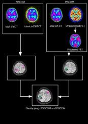
This study analyzed SOZ localization in 37 patients using SISCOM and PISCOM, corroborated by postoperative MRI and outcome. We aimed to determine optimal z-scores for both methods and investigated the influence of the SPECT injection time-seizure duration ratio on interpretation of these maps in SOZ localization accuracy. We also created a ratio image representing the overlap between SISCOM and PISCOM maps, using Z-scores greater than 1, 1.5, and 2 for both maps.
Results
Of 21 Engel-1 patients, in 16 (76 %) the SISCOM and PISCOM perfusion (large, high intensity) lateralized to the resection side, in 3 (14 %) patients to the contralateral side, and in 2 (10 %) showed bilateral activation. In Engel 1 patients, hyperperfusion on the PISCOM mapped the resection zone in 90 % of cases, compared to 80 % with the SISCOM map. The optimal z-scores were 1.9 for SISCOM, yielding a sensitivity of 88.5 % and specificity of 94.8 % for mapping the SOZ. For PISCOM, the optimal z-score was 2.2, achieving a sensitivity of 87.4 % and specificity of 93.3 %. The ratio map, created from SISCOM and PISCOM overlap (z-scores > 2), was useful for delineating the SOZ and aligned better with the resection cavity than SISCOM and PISCOM maps. The analysis showed no significant median differences in injection-time-to-seizure-duration ratios between seizure-free and non-seizure-free patients (p = 0.8). We found a difference in the median injection-time-to-seizure-duration ratio between cases where SISCOM/PISCOM maps were concordant with the resection cavity in seizure-free patients, at 0.09 (0.06, 0.28), and discordant cases, at 0.41 (0.17, 0.61).
Conclusion
This study determined the optimal Z-score for mapping the SOZ and evaluated the use of ratio image in SOZ localization. Both SISCOM and PISCOM demonstrated their utility in identifying the SOZ in epilepsy surgery patients, with PISCOM emerging as a promising alternative to SISCOM, potentially eliminating the need for interictal SPECT.
评估SISCOM和PISCOM在癫痫手术中癫痫区定位中的效用:一项基于结果的比较研究
内源性SPECT成像在非侵入性工具中脱颖而出,成为确定癫痫网络空间的主要手术前方法。先进的图像分析方法如SISCOM和PISCOM提高了SPECT图像的解译能力。然而,关于它们的比较效用、重叠区域的重要性以及映射/定位癫痫发作区域的最佳z分数的研究是有限的。因此,本研究集中于对这些图像进行详细分析,绘制脑电图网络和定位癫痫发作区。方法本研究分析了37例使用SISCOM和PISCOM的患者的SOZ定位,并通过术后MRI和预后进行证实。我们的目标是确定两种方法的最佳z分数,并研究SPECT注射时间-癫痫持续时间比对SOZ定位精度解释这些图谱的影响。我们还创建了一个比率图像,表示SISCOM和PISCOM地图之间的重叠,使用大于1、1.5和2的z分数来表示这两个地图。结果在21例Engel-1患者中,16例(76%)患者的SISCOM和PISCOM灌注(大、高强度)偏向切除侧,3例(14%)患者偏向对侧,2例(10%)患者双侧激活。在Engel 1患者中,90%的病例在PISCOM上的高灌注映射出切除区,而在SISCOM上的高灌注映射为80%。SISCOM的最佳z得分为1.9,对SOZ的定位敏感性为88.5%,特异性为94.8%。PISCOM的最佳z-score为2.2,灵敏度为87.4%,特异性为93.3%。由SISCOM和PISCOM重叠(z-scores > 2)创建的比率图有助于描绘SOZ,并且比SISCOM和PISCOM地图与切除腔更好地对齐。分析显示,无癫痫发作和非无癫痫发作患者的注射时间与癫痫发作持续时间比值中位数无显著差异(p = 0.8)。我们发现,SISCOM/PISCOM图谱与无癫痫患者切除腔一致的病例中,注射时间与癫痫持续时间的中位数比为0.09(0.06,0.28),而不一致的病例中,注射时间与癫痫持续时间的中位数比为0.41(0.17,0.61)。结论确定了SOZ定位的最佳z值,并评价了比值图像在SOZ定位中的应用。SISCOM和PISCOM都证明了它们在癫痫手术患者中识别SOZ的实用性,PISCOM成为SISCOM的一个有希望的替代方案,有可能消除对间期SPECT的需求。
本文章由计算机程序翻译,如有差异,请以英文原文为准。
求助全文
约1分钟内获得全文
求助全文
来源期刊

Journal of Clinical Neuroscience
医学-临床神经学
CiteScore
4.50
自引率
0.00%
发文量
402
审稿时长
40 days
期刊介绍:
This International journal, Journal of Clinical Neuroscience, publishes articles on clinical neurosurgery and neurology and the related neurosciences such as neuro-pathology, neuro-radiology, neuro-ophthalmology and neuro-physiology.
The journal has a broad International perspective, and emphasises the advances occurring in Asia, the Pacific Rim region, Europe and North America. The Journal acts as a focus for publication of major clinical and laboratory research, as well as publishing solicited manuscripts on specific subjects from experts, case reports and other information of interest to clinicians working in the clinical neurosciences.
 求助内容:
求助内容: 应助结果提醒方式:


