Masihuz Zaman, Shu Yang, Ya Huang, Jay M. Yarbro, Yanhong Hao, Zhen Wang, Danting Liu, Kiara E. Harper, Hadeer Soliman, Alex Hemphill, Sarah Harvey, Shondra M. Pruett-Miller, Valerie Stewart, Ajay Singh Tanwar, Ravi Kalathur, Christy R. Grace, Martin Turk, Sagar Chittori, Yun Jiao, Zhiping Wu, Anthony A. High, Xusheng Wang, Geidy E. Serrano, Thomas G. Beach, Gang Yu, Yang Yang, Ping-Chung Chen, Junmin Peng
{"title":"Midkine attenuates amyloid-β fibril assembly and plaque formation","authors":"Masihuz Zaman, Shu Yang, Ya Huang, Jay M. Yarbro, Yanhong Hao, Zhen Wang, Danting Liu, Kiara E. Harper, Hadeer Soliman, Alex Hemphill, Sarah Harvey, Shondra M. Pruett-Miller, Valerie Stewart, Ajay Singh Tanwar, Ravi Kalathur, Christy R. Grace, Martin Turk, Sagar Chittori, Yun Jiao, Zhiping Wu, Anthony A. High, Xusheng Wang, Geidy E. Serrano, Thomas G. Beach, Gang Yu, Yang Yang, Ping-Chung Chen, Junmin Peng","doi":"10.1038/s41594-025-01657-8","DOIUrl":null,"url":null,"abstract":"<p>Proteomic profiling of Alzheimer disease (AD) brains has identified numerous understudied proteins, including midkine (MDK), that are highly upregulated and correlated with amyloid-β (Aβ) from the early disease stage but their roles in disease progression are not fully understood. Here, we present that MDK attenuates Aβ assembly and influences amyloid formation in the 5xFAD amyloidosis mouse model. MDK protein mitigates fibril formation of both Aβ40 and Aβ42 peptides according to thioflavin T fluorescence, circular dichroism, negative-stain electron microscopy and nuclear magnetic resonance analyses. Knockout of the <i>Mdk</i> gene in 5xFAD increased amyloid formation and microglial activation in the brain. Further comprehensive mass-spectrometry-based profiling of the whole proteome and detergent-insoluble proteome in these mouse models indicated significant accumulation of Aβ and Aβ-correlated proteins, along with microglial components. Thus, our structural and mouse model studies reveal a protective role of MDK in counteracting amyloid pathology in AD.</p>","PeriodicalId":18822,"journal":{"name":"Nature structural & molecular biology","volume":"22 1","pages":""},"PeriodicalIF":0.0000,"publicationDate":"2025-08-21","publicationTypes":"Journal Article","fieldsOfStudy":null,"isOpenAccess":false,"openAccessPdf":"","citationCount":"0","resultStr":null,"platform":"Semanticscholar","paperid":null,"PeriodicalName":"Nature structural & molecular biology","FirstCategoryId":"1085","ListUrlMain":"https://doi.org/10.1038/s41594-025-01657-8","RegionNum":0,"RegionCategory":null,"ArticlePicture":[],"TitleCN":null,"AbstractTextCN":null,"PMCID":null,"EPubDate":"","PubModel":"","JCR":"","JCRName":"","Score":null,"Total":0}
引用次数: 0
Abstract
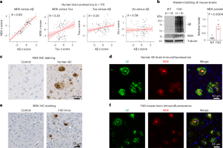
Proteomic profiling of Alzheimer disease (AD) brains has identified numerous understudied proteins, including midkine (MDK), that are highly upregulated and correlated with amyloid-β (Aβ) from the early disease stage but their roles in disease progression are not fully understood. Here, we present that MDK attenuates Aβ assembly and influences amyloid formation in the 5xFAD amyloidosis mouse model. MDK protein mitigates fibril formation of both Aβ40 and Aβ42 peptides according to thioflavin T fluorescence, circular dichroism, negative-stain electron microscopy and nuclear magnetic resonance analyses. Knockout of the Mdk gene in 5xFAD increased amyloid formation and microglial activation in the brain. Further comprehensive mass-spectrometry-based profiling of the whole proteome and detergent-insoluble proteome in these mouse models indicated significant accumulation of Aβ and Aβ-correlated proteins, along with microglial components. Thus, our structural and mouse model studies reveal a protective role of MDK in counteracting amyloid pathology in AD.

 求助内容:
求助内容: 应助结果提醒方式:


