Assessing the effects of luminescently labelled and non-labelled PET nanoparticles on environmental bacteria
IF 8.1
2区 环境科学与生态学
Q1 ENVIRONMENTAL SCIENCES
引用次数: 0
Abstract
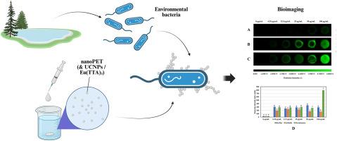
Manufacturers use polyethylene terephthalate (PET) to create many everyday objects, which break down into nanoparticles when released into the environment. This fact raises questions about the effects of nanometric PET on living organisms, including bacteria. However, studies on nanoPET are rare and challenging, even if only because its detection and visualisation are difficult. We studied nanoPET toxicity on selected bacteria and tested its visualisation in biological samples using three nanoPET types: pure, labelled with upconverting nanoparticles (UCNPs), and labelled with Eu3+ complex compound. The resulting colloids were characterized using dynamic light scattering, zeta potential, and emission measurements. The results confirmed the development of a method for preparing a stable aqueous colloid of nanoPET. Toxicity tests on Bacillus, Lelliottia, and Pseudomonas strains were carried out using the AlamarBlue method, along with measurements of Glutathione S-Transferase enzyme activity. The impact of labelled and non-labelled nanoPET on bacterial carbon utilisation of different sources was also evaluated. Crystal violet and the o-nitrophenyl-β-d-galactoside assays were applied to assess changes in membrane permeability. The adhesion of nanoPET to bacterial cells was examined using atomic force microscopy, and biofilm alterations were visualised under an optical microscope. UCNPs enabled the detection of nanoPET aggregation in bacterial biofilms. PET nanoparticles had a neutral or stimulating effect on bacterial growth. Cell membrane permeability varied depending on the bacterial strain and the type of nanoPET used. The results offer valuable insight into the environmental impact of nanoPET and demonstrate the effectiveness of the new nanoplastic labelling and detection method.
评估发光标记和未标记的PET纳米颗粒对环境细菌的影响
制造商使用聚对苯二甲酸乙二醇酯(PET)制造许多日常用品,这些物品被释放到环境中时会分解成纳米颗粒。这一事实引发了关于纳米PET对包括细菌在内的生物体的影响的问题。然而,纳米et的研究是罕见的和具有挑战性的,即使只是因为它的检测和可视化是困难的。我们研究了纳米et对选定细菌的毒性,并使用三种类型的纳米et测试其在生物样品中的可视化效果:纯纳米et、上转化纳米颗粒(UCNPs)标记和Eu3+复合物标记。所得到的胶体用动态光散射、zeta电位和发射测量来表征。结果证实了制备稳定的纳米et水凝胶的方法的发展。采用AlamarBlue方法对芽孢杆菌、小叶菌和假单胞菌菌株进行毒性试验,并测定谷胱甘肽s -转移酶活性。还评估了标记和未标记的纳米et对不同来源的细菌碳利用的影响。采用结晶紫法和邻硝基苯-β-d-半乳糖苷法评价膜通透性的变化。使用原子力显微镜检查纳米et与细菌细胞的粘附,并在光学显微镜下观察生物膜的变化。UCNPs可以检测细菌生物膜中的纳米et聚集。PET纳米颗粒对细菌生长具有中性或刺激作用。细胞膜通透性取决于细菌菌株和使用的纳米et类型。这些结果为纳米塑料对环境的影响提供了有价值的见解,并证明了新的纳米塑料标记和检测方法的有效性。
本文章由计算机程序翻译,如有差异,请以英文原文为准。
求助全文
约1分钟内获得全文
求助全文
来源期刊
Chemosphere
环境科学-环境科学
CiteScore
15.80
自引率
8.00%
发文量
4975
审稿时长
3.4 months
期刊介绍:
Chemosphere, being an international multidisciplinary journal, is dedicated to publishing original communications and review articles on chemicals in the environment. The scope covers a wide range of topics, including the identification, quantification, behavior, fate, toxicology, treatment, and remediation of chemicals in the bio-, hydro-, litho-, and atmosphere, ensuring the broad dissemination of research in this field.
 求助内容:
求助内容: 应助结果提醒方式:


