Xu Wang, Patrice Monkam, Bonan Zhao, Shouliang Qi, He Ma, Long Huang, Wei Qian
{"title":"Enhancing Lesion Segmentation in Ultrasound Images: The Impact of Targeted Data Augmentation Strategies.","authors":"Xu Wang, Patrice Monkam, Bonan Zhao, Shouliang Qi, He Ma, Long Huang, Wei Qian","doi":"10.1155/ijbi/3309822","DOIUrl":null,"url":null,"abstract":"<p><p>Automated lesion segmentation in ultrasound (US) images based on deep learning (DL) approaches plays a crucial role in disease diagnosis and treatment. However, the successful implementation of these approaches is conditioned by large-scale and diverse annotated datasets whose obtention is tedious and expertise demanding. Although methods like generative adversarial networks (GANs) can help address sample scarcity, they are often associated with complex training processes and high computational demands, which can limit their practicality and feasibility, especially in resource-constrained scenarios. Therefore, this study is aimed at exploring new solutions to address the challenge of limited annotated samples in automated lesion delineation in US images. Specifically, we propose five distinct mixed sample augmentation strategies and assess their effectiveness using four deep segmentation models for the delineation of two lesion types: breast and thyroid lesions. Extensive experimental analyses indicate that the effectiveness of these augmentation strategies is strongly influenced by both the lesion type and the model architecture. When appropriately selected, these strategies result in substantial performance improvements, with the Dice and Jaccard indices increasing by up to 37.95% and 36.32% for breast lesions and 14.59% and 13.01% for thyroid lesions, respectively. These improvements highlight the potential of the proposed strategies as a reliable solution to address data scarcity in automated lesion segmentation tasks. Furthermore, the study emphasizes the critical importance of carefully selecting data augmentation approaches, offering valuable insights into how their strategic application can significantly enhance the performance of DL models.</p>","PeriodicalId":47063,"journal":{"name":"International Journal of Biomedical Imaging","volume":"2025 ","pages":"3309822"},"PeriodicalIF":1.3000,"publicationDate":"2025-08-11","publicationTypes":"Journal Article","fieldsOfStudy":null,"isOpenAccess":false,"openAccessPdf":"https://www.ncbi.nlm.nih.gov/pmc/articles/PMC12360876/pdf/","citationCount":"0","resultStr":null,"platform":"Semanticscholar","paperid":null,"PeriodicalName":"International Journal of Biomedical Imaging","FirstCategoryId":"1085","ListUrlMain":"https://doi.org/10.1155/ijbi/3309822","RegionNum":0,"RegionCategory":null,"ArticlePicture":[],"TitleCN":null,"AbstractTextCN":null,"PMCID":null,"EPubDate":"2025/1/1 0:00:00","PubModel":"eCollection","JCR":"Q2","JCRName":"ENGINEERING, BIOMEDICAL","Score":null,"Total":0}
引用次数: 0
Abstract
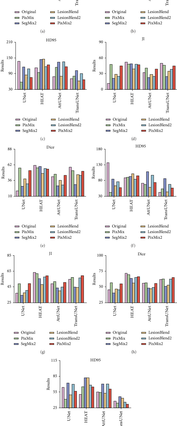
Automated lesion segmentation in ultrasound (US) images based on deep learning (DL) approaches plays a crucial role in disease diagnosis and treatment. However, the successful implementation of these approaches is conditioned by large-scale and diverse annotated datasets whose obtention is tedious and expertise demanding. Although methods like generative adversarial networks (GANs) can help address sample scarcity, they are often associated with complex training processes and high computational demands, which can limit their practicality and feasibility, especially in resource-constrained scenarios. Therefore, this study is aimed at exploring new solutions to address the challenge of limited annotated samples in automated lesion delineation in US images. Specifically, we propose five distinct mixed sample augmentation strategies and assess their effectiveness using four deep segmentation models for the delineation of two lesion types: breast and thyroid lesions. Extensive experimental analyses indicate that the effectiveness of these augmentation strategies is strongly influenced by both the lesion type and the model architecture. When appropriately selected, these strategies result in substantial performance improvements, with the Dice and Jaccard indices increasing by up to 37.95% and 36.32% for breast lesions and 14.59% and 13.01% for thyroid lesions, respectively. These improvements highlight the potential of the proposed strategies as a reliable solution to address data scarcity in automated lesion segmentation tasks. Furthermore, the study emphasizes the critical importance of carefully selecting data augmentation approaches, offering valuable insights into how their strategic application can significantly enhance the performance of DL models.
期刊介绍:
The International Journal of Biomedical Imaging is managed by a board of editors comprising internationally renowned active researchers. The journal is freely accessible online and also offered for purchase in print format. It employs a web-based review system to ensure swift turnaround times while maintaining high standards. In addition to regular issues, special issues are organized by guest editors. The subject areas covered include (but are not limited to):
Digital radiography and tomosynthesis
X-ray computed tomography (CT)
Magnetic resonance imaging (MRI)
Single photon emission computed tomography (SPECT)
Positron emission tomography (PET)
Ultrasound imaging
Diffuse optical tomography, coherence, fluorescence, bioluminescence tomography, impedance tomography
Neutron imaging for biomedical applications
Magnetic and optical spectroscopy, and optical biopsy
Optical, electron, scanning tunneling/atomic force microscopy
Small animal imaging
Functional, cellular, and molecular imaging
Imaging assays for screening and molecular analysis
Microarray image analysis and bioinformatics
Emerging biomedical imaging techniques
Imaging modality fusion
Biomedical imaging instrumentation
Biomedical image processing, pattern recognition, and analysis
Biomedical image visualization, compression, transmission, and storage
Imaging and modeling related to systems biology and systems biomedicine
Applied mathematics, applied physics, and chemistry related to biomedical imaging
Grid-enabling technology for biomedical imaging and informatics

 求助内容:
求助内容: 应助结果提醒方式:


