Sabīne Teifurova, Kārlis Rācenis, Ģirts Freijs, Sigita Skrastina, Arturs Balodis
{"title":"Radiological Findings of Retrograde Venous Cerebral Air Embolism Infarcts: A Case Report and Literature Review.","authors":"Sabīne Teifurova, Kārlis Rācenis, Ģirts Freijs, Sigita Skrastina, Arturs Balodis","doi":"10.2147/VHRM.S537865","DOIUrl":null,"url":null,"abstract":"<p><strong>Background: </strong>Cerebral air embolism infarction (CAEI) is a rare but life-threatening condition that can affect the venous or arterial blood supply to the brain. Its aetiology is mostly iatrogenic, often resulting from complications of cardiothoracic or neurosurgical procedures, as well as manipulations with peripheral or central catheters. If undiagnosed and untreated, cerebral air embolism infarction can lead to long-term neurological consequences or even death. Diagnosis relies on clinical presentation and neuroimaging findings from CT and MRI, which are time-sensitive and not well described in the current literature.</p><p><strong>Case presentation: </strong>We present a rare case of cerebral infarction as a complication of retrograde cerebral venous air embolism following haemodialysis catheter removal in a young patient, with management leading to a favourable outcome. The diagnosis was confirmed based on clinical manifestations and neuroimaging findings, with air emboli identified in the subarachnoid space on the CT scan, followed by characteristic MRI changes defined for cerebral air embolism infarcts. Timely diagnosis allowed for the rapid initiation of hyperbaric oxygen therapy and the rehabilitation process, resulting in positive outcomes.</p><p><strong>Conclusion: </strong>Timely neuroimaging-particularly CT within the first 2 hours-is critical for diagnosing CAEI. MRI findings, including cytotoxic and vasogenic oedema in a distal vascular distribution and leptomeningeal enhancement, further support diagnosis. Early identification and treatment initiation are essential for improving patient outcomes.</p>","PeriodicalId":23597,"journal":{"name":"Vascular Health and Risk Management","volume":"21 ","pages":"617-631"},"PeriodicalIF":2.8000,"publicationDate":"2025-08-12","publicationTypes":"Journal Article","fieldsOfStudy":null,"isOpenAccess":false,"openAccessPdf":"https://www.ncbi.nlm.nih.gov/pmc/articles/PMC12358500/pdf/","citationCount":"0","resultStr":null,"platform":"Semanticscholar","paperid":null,"PeriodicalName":"Vascular Health and Risk Management","FirstCategoryId":"1085","ListUrlMain":"https://doi.org/10.2147/VHRM.S537865","RegionNum":0,"RegionCategory":null,"ArticlePicture":[],"TitleCN":null,"AbstractTextCN":null,"PMCID":null,"EPubDate":"2025/1/1 0:00:00","PubModel":"eCollection","JCR":"Q2","JCRName":"PERIPHERAL VASCULAR DISEASE","Score":null,"Total":0}
引用次数: 0
Abstract
Background: Cerebral air embolism infarction (CAEI) is a rare but life-threatening condition that can affect the venous or arterial blood supply to the brain. Its aetiology is mostly iatrogenic, often resulting from complications of cardiothoracic or neurosurgical procedures, as well as manipulations with peripheral or central catheters. If undiagnosed and untreated, cerebral air embolism infarction can lead to long-term neurological consequences or even death. Diagnosis relies on clinical presentation and neuroimaging findings from CT and MRI, which are time-sensitive and not well described in the current literature.
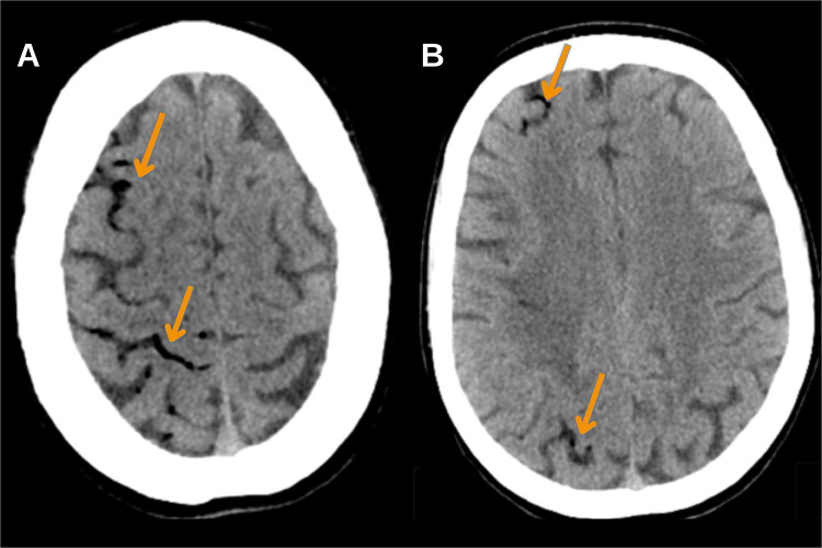
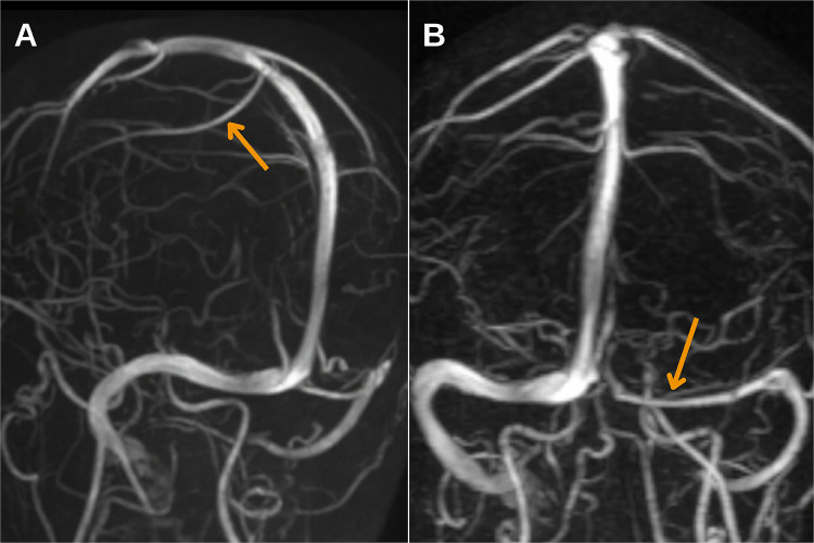
Case presentation: We present a rare case of cerebral infarction as a complication of retrograde cerebral venous air embolism following haemodialysis catheter removal in a young patient, with management leading to a favourable outcome. The diagnosis was confirmed based on clinical manifestations and neuroimaging findings, with air emboli identified in the subarachnoid space on the CT scan, followed by characteristic MRI changes defined for cerebral air embolism infarcts. Timely diagnosis allowed for the rapid initiation of hyperbaric oxygen therapy and the rehabilitation process, resulting in positive outcomes.
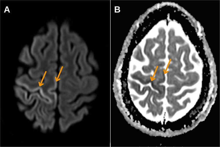
Conclusion: Timely neuroimaging-particularly CT within the first 2 hours-is critical for diagnosing CAEI. MRI findings, including cytotoxic and vasogenic oedema in a distal vascular distribution and leptomeningeal enhancement, further support diagnosis. Early identification and treatment initiation are essential for improving patient outcomes.
期刊介绍:
An international, peer-reviewed journal of therapeutics and risk management, focusing on concise rapid reporting of clinical studies on the processes involved in the maintenance of vascular health; the monitoring, prevention, and treatment of vascular disease and its sequelae; and the involvement of metabolic disorders, particularly diabetes. In addition, the journal will also seek to define drug usage in terms of ultimate uptake and acceptance by the patient and healthcare professional.

 求助内容:
求助内容: 应助结果提醒方式:


