Improving risk stratification of PI-RADS 3 + 1 lesions of the peripheral zone: expert lexicon of terms, multi-reader performance and contribution of artificial intelligence.
Philip A Glemser, Nils Netzer, Christian H Ziener, Markus Wilhelm, Thomas Hielscher, Kevin Sun Zhang, Magdalena Görtz, Viktoria Schütz, Albrecht Stenzinger, Markus Hohenfellner, Heinz-Peter Schlemmer, David Bonekamp
{"title":"Improving risk stratification of PI-RADS 3 + 1 lesions of the peripheral zone: expert lexicon of terms, multi-reader performance and contribution of artificial intelligence.","authors":"Philip A Glemser, Nils Netzer, Christian H Ziener, Markus Wilhelm, Thomas Hielscher, Kevin Sun Zhang, Magdalena Görtz, Viktoria Schütz, Albrecht Stenzinger, Markus Hohenfellner, Heinz-Peter Schlemmer, David Bonekamp","doi":"10.1186/s40644-025-00916-7","DOIUrl":null,"url":null,"abstract":"<p><strong>Background: </strong>According to PI-RADS v2.1, peripheral PI-RADS 3 lesions are upgraded to PI-RADS 4 if dynamic contrast-enhanced MRI is positive (3+1 lesions), however those lesions are radiologically challenging. We aimed to define criteria by expert consensus and test applicability by other radiologists for sPC prediction of PI-RADS 3+1 lesions and determine their value in integrated regression models.</p><p><strong>Methods: </strong>From consecutive 3 Tesla MR examinations performed between 08/2016 to 12/2018 we identified 85 MRI examinations from 83 patients with a total of 94 PI-RADS 3+1 lesions in the official clinical report. Lesions were retrospectively assessed by expert consensus with construction of a newly devised feature catalogue which was utilized subsequently by two additional radiologists specialized in prostate MRI for independent lesion assessment. With reference to extended fused targeted and systematic TRUS/MRI-biopsy histopathological correlation, relevant catalogue features were identified by univariate analysis and put into context to typically available clinical features and automated AI image assessment utilizing lasso-penalized logistic regression models, also focusing on the contribution of DCE imaging (feature-based, bi- and multiparametric AI-enhanced and solely bi- and multiparametric AI-driven).</p><p><strong>Results: </strong>The feature catalog enabled image-based lesional risk stratification for all readers. Expert consensus provided 3 significant features in univariate analysis (adj. p-value <0.05; most relevant feature T2w configuration: \"irregular/microlobulated/spiculated\", OR 9.0 (95%CI 2.3-44.3); adj. p-value: 0.016). These remained after lasso penalized regression based feature reduction, while the only selected clinical feature was prostate volume (OR<1), enabling nomogram construction. While DCE-derived consensus features did not enhance model performance (bootstrapped AUC), there was a trend for increased performance by including multiparametric AI, but not biparametric AI into models, both for combined and AI-only models.</p><p><strong>Conclusions: </strong>PI-RADS 3+1 lesions can be risk-stratified using lexicon terms and a key feature nomogram. AI potentially benefits more from DCE imaging than experienced prostate radiologists.</p><p><strong>Clinical trial number: </strong>Not applicable.</p>","PeriodicalId":9548,"journal":{"name":"Cancer Imaging","volume":"25 1","pages":"102"},"PeriodicalIF":3.5000,"publicationDate":"2025-08-19","publicationTypes":"Journal Article","fieldsOfStudy":null,"isOpenAccess":false,"openAccessPdf":"https://www.ncbi.nlm.nih.gov/pmc/articles/PMC12366217/pdf/","citationCount":"0","resultStr":null,"platform":"Semanticscholar","paperid":null,"PeriodicalName":"Cancer Imaging","FirstCategoryId":"3","ListUrlMain":"https://doi.org/10.1186/s40644-025-00916-7","RegionNum":2,"RegionCategory":"医学","ArticlePicture":[],"TitleCN":null,"AbstractTextCN":null,"PMCID":null,"EPubDate":"","PubModel":"","JCR":"Q2","JCRName":"ONCOLOGY","Score":null,"Total":0}
引用次数: 0
Abstract
Background: According to PI-RADS v2.1, peripheral PI-RADS 3 lesions are upgraded to PI-RADS 4 if dynamic contrast-enhanced MRI is positive (3+1 lesions), however those lesions are radiologically challenging. We aimed to define criteria by expert consensus and test applicability by other radiologists for sPC prediction of PI-RADS 3+1 lesions and determine their value in integrated regression models.
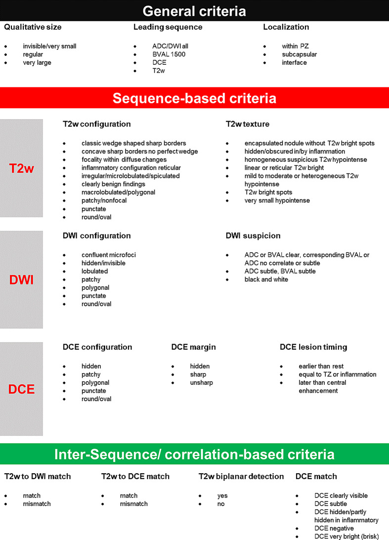
Methods: From consecutive 3 Tesla MR examinations performed between 08/2016 to 12/2018 we identified 85 MRI examinations from 83 patients with a total of 94 PI-RADS 3+1 lesions in the official clinical report. Lesions were retrospectively assessed by expert consensus with construction of a newly devised feature catalogue which was utilized subsequently by two additional radiologists specialized in prostate MRI for independent lesion assessment. With reference to extended fused targeted and systematic TRUS/MRI-biopsy histopathological correlation, relevant catalogue features were identified by univariate analysis and put into context to typically available clinical features and automated AI image assessment utilizing lasso-penalized logistic regression models, also focusing on the contribution of DCE imaging (feature-based, bi- and multiparametric AI-enhanced and solely bi- and multiparametric AI-driven).
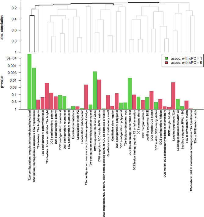
Results: The feature catalog enabled image-based lesional risk stratification for all readers. Expert consensus provided 3 significant features in univariate analysis (adj. p-value <0.05; most relevant feature T2w configuration: "irregular/microlobulated/spiculated", OR 9.0 (95%CI 2.3-44.3); adj. p-value: 0.016). These remained after lasso penalized regression based feature reduction, while the only selected clinical feature was prostate volume (OR<1), enabling nomogram construction. While DCE-derived consensus features did not enhance model performance (bootstrapped AUC), there was a trend for increased performance by including multiparametric AI, but not biparametric AI into models, both for combined and AI-only models.
Conclusions: PI-RADS 3+1 lesions can be risk-stratified using lexicon terms and a key feature nomogram. AI potentially benefits more from DCE imaging than experienced prostate radiologists.
Cancer ImagingONCOLOGY-RADIOLOGY, NUCLEAR MEDICINE & MEDICAL IMAGING
CiteScore
7.00
自引率
0.00%
发文量
66
审稿时长
>12 weeks
期刊介绍:
Cancer Imaging is an open access, peer-reviewed journal publishing original articles, reviews and editorials written by expert international radiologists working in oncology.
The journal encompasses CT, MR, PET, ultrasound, radionuclide and multimodal imaging in all kinds of malignant tumours, plus new developments, techniques and innovations. Topics of interest include:
Breast Imaging
Chest
Complications of treatment
Ear, Nose & Throat
Gastrointestinal
Hepatobiliary & Pancreatic
Imaging biomarkers
Interventional
Lymphoma
Measurement of tumour response
Molecular functional imaging
Musculoskeletal
Neuro oncology
Nuclear Medicine
Paediatric.


 求助内容:
求助内容: 应助结果提醒方式:


