Eric V Mastrolonardo, Sarah Sussman, Bo Yun, Victor Jegede, Dev R Amin, Joel Rosenbloom, Andrew P South, Voichita Bar-Ad, Peter J Wermuth, Adam J Luginbuhl
{"title":"Effects of Oxygen Manipulation on Myofibroblast Phenotypic Transformation in Patients With Radiation-Induced Fibrosis.","authors":"Eric V Mastrolonardo, Sarah Sussman, Bo Yun, Victor Jegede, Dev R Amin, Joel Rosenbloom, Andrew P South, Voichita Bar-Ad, Peter J Wermuth, Adam J Luginbuhl","doi":"10.1111/wrr.70075","DOIUrl":null,"url":null,"abstract":"<p><p>We tested if hyperoxic conditions can reduce the proportion of active myofibroblasts, which are assumed to be a major driver of head and neck radiation-induced fibrosis, as measured by expression levels of pro-fibrotic genes. Radiated, non-cancerous soft tissue from the head and neck and skin/soft tissue from non-radiated flap donor site were collected from each patient. Myofibroblast density was quantified using immunofluorescence staining with α-SMA and DAPI and visualisation under confocal microscopy and compared between baseline non-radiated and radiated tissue from the same patient. From each tissue specimen, fibroblast cell lines were cultured and exposed to either normoxic, hypoxic, or hyperoxic conditions for 10 days. Total RNA was extracted and reverse-transcribed, and gene expression levels were quantified using RT-PCR. Relative gene expression levels of pro-fibrotic genes COL1A1, COL3A1, FN-EDA, α-SMA, HIF-1α, VEGFα, and VEGFR were compared between normoxic, hypoxic, and hyperoxic treatment groups. Three patients with six total tissue samples were acquired. Radiated tissue contained a higher density of myofibroblasts (calculated as cells/mm<sup>2</sup>) and demonstrated higher expression of pro-fibrotic genes than non-radiated donor site tissue. Hyperoxia decreases expression levels of pro-fibrotic genes in radiated and non-radiated tissue, while hypoxia increases pro-fibrotic gene expression levels in radiated and non-radiated tissue. Study findings indicate that hypoxia is a driver of myofibroblast activation and that subjects with radiation-induced fibrosis of the head and neck have increased expression of myofibroblastic phenotype. Hyperoxygenation can reduce the proportion of active myofibroblasts, revealing a potential therapeutic method to halt chronic fibrotic pathways.</p>","PeriodicalId":23864,"journal":{"name":"Wound Repair and Regeneration","volume":"33 4","pages":"e70075"},"PeriodicalIF":3.4000,"publicationDate":"2025-07-01","publicationTypes":"Journal Article","fieldsOfStudy":null,"isOpenAccess":false,"openAccessPdf":"https://www.ncbi.nlm.nih.gov/pmc/articles/PMC12358765/pdf/","citationCount":"0","resultStr":null,"platform":"Semanticscholar","paperid":null,"PeriodicalName":"Wound Repair and Regeneration","FirstCategoryId":"3","ListUrlMain":"https://doi.org/10.1111/wrr.70075","RegionNum":3,"RegionCategory":"医学","ArticlePicture":[],"TitleCN":null,"AbstractTextCN":null,"PMCID":null,"EPubDate":"","PubModel":"","JCR":"Q2","JCRName":"CELL BIOLOGY","Score":null,"Total":0}
引用次数: 0
Abstract
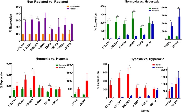
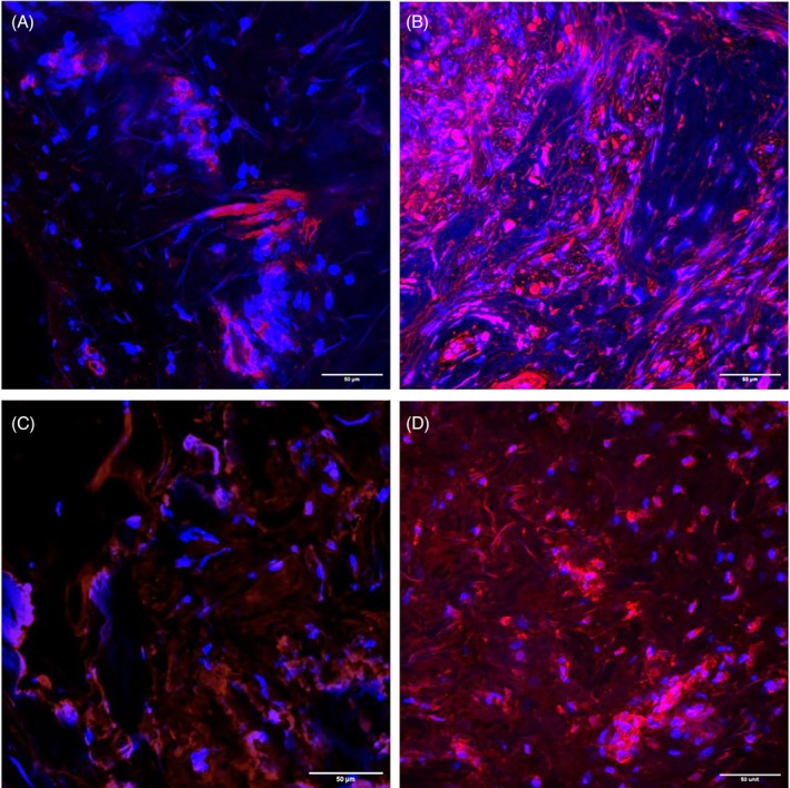
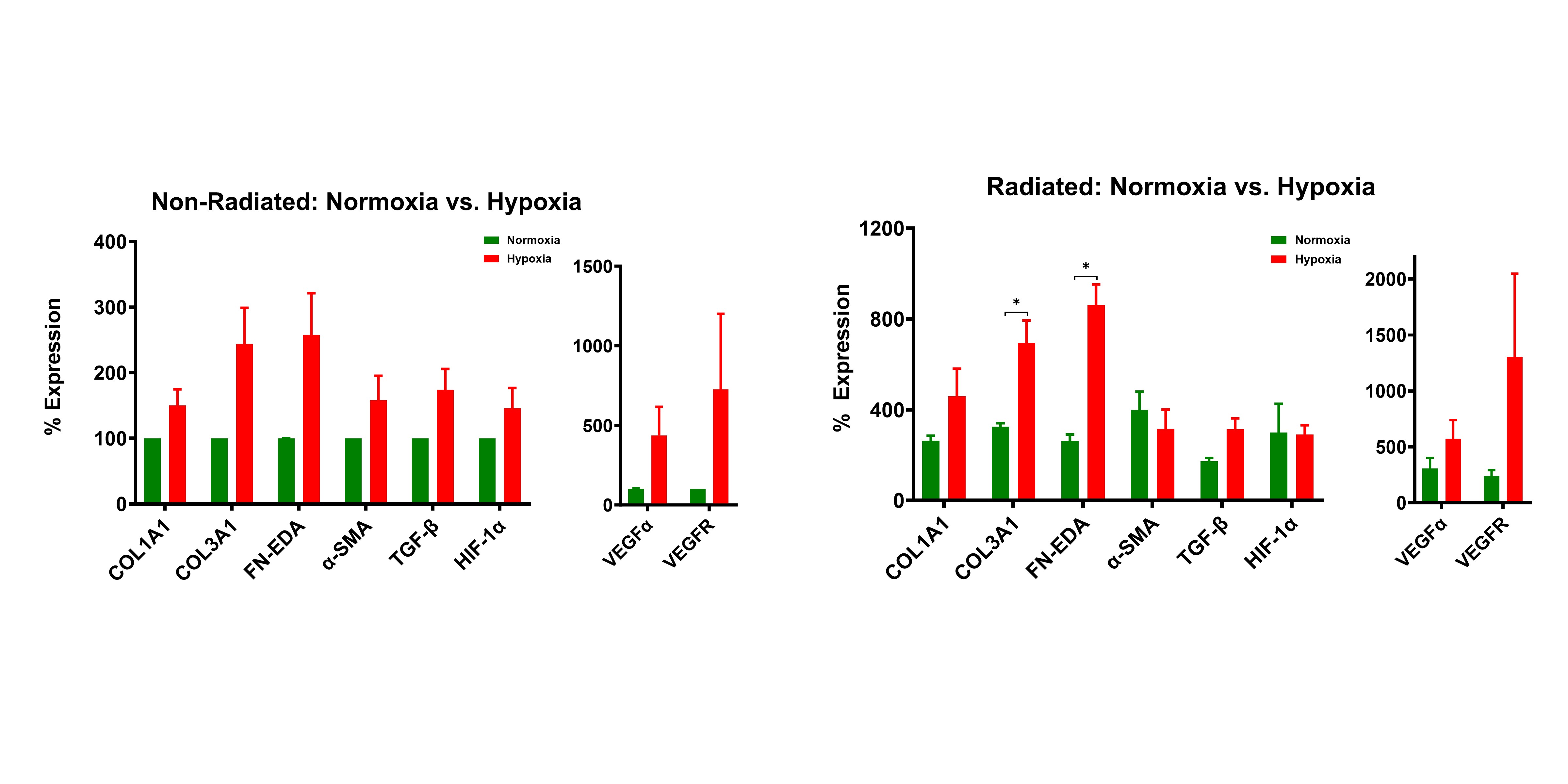
We tested if hyperoxic conditions can reduce the proportion of active myofibroblasts, which are assumed to be a major driver of head and neck radiation-induced fibrosis, as measured by expression levels of pro-fibrotic genes. Radiated, non-cancerous soft tissue from the head and neck and skin/soft tissue from non-radiated flap donor site were collected from each patient. Myofibroblast density was quantified using immunofluorescence staining with α-SMA and DAPI and visualisation under confocal microscopy and compared between baseline non-radiated and radiated tissue from the same patient. From each tissue specimen, fibroblast cell lines were cultured and exposed to either normoxic, hypoxic, or hyperoxic conditions for 10 days. Total RNA was extracted and reverse-transcribed, and gene expression levels were quantified using RT-PCR. Relative gene expression levels of pro-fibrotic genes COL1A1, COL3A1, FN-EDA, α-SMA, HIF-1α, VEGFα, and VEGFR were compared between normoxic, hypoxic, and hyperoxic treatment groups. Three patients with six total tissue samples were acquired. Radiated tissue contained a higher density of myofibroblasts (calculated as cells/mm2) and demonstrated higher expression of pro-fibrotic genes than non-radiated donor site tissue. Hyperoxia decreases expression levels of pro-fibrotic genes in radiated and non-radiated tissue, while hypoxia increases pro-fibrotic gene expression levels in radiated and non-radiated tissue. Study findings indicate that hypoxia is a driver of myofibroblast activation and that subjects with radiation-induced fibrosis of the head and neck have increased expression of myofibroblastic phenotype. Hyperoxygenation can reduce the proportion of active myofibroblasts, revealing a potential therapeutic method to halt chronic fibrotic pathways.
期刊介绍:
Wound Repair and Regeneration provides extensive international coverage of cellular and molecular biology, connective tissue, and biological mediator studies in the field of tissue repair and regeneration and serves a diverse audience of surgeons, plastic surgeons, dermatologists, biochemists, cell biologists, and others.
Wound Repair and Regeneration is the official journal of The Wound Healing Society, The European Tissue Repair Society, The Japanese Society for Wound Healing, and The Australian Wound Management Association.
 求助内容:
求助内容: 应助结果提醒方式:


