{"title":"Innovative Biomarkers for Diagnosing Malignant Ascites in Liver Cancer.","authors":"Yan Zhang, Jing Wu, Huaizhong Cui, Xiaojing Zhang, Lingyan He, Kailong Gu, Aifang Xu","doi":"10.2147/OTT.S527224","DOIUrl":null,"url":null,"abstract":"<p><strong>Background: </strong>Liver cancer ranks among the most prevalent and lethal malignancies worldwide, with metastatic malignant ascites being a common complication. This study seeks to assess the diagnostic significance of high fluorescent cells (HFCs), biochemical and tumor markers in predicting the development of metastatic malignant ascites in patients with liver cancer.</p><p><strong>Methods: </strong>We collected ascites samples from 266 patients diagnosed with liver cancer. HFC were analyzed using the BF mode of the BC-7500 hematology analyzer, assessing both relative counts (HF-BF%) and absolute counts (HF-BF#). Additionally, biochemical and tumor markers were evaluated in serum and ascites. The diagnostic accuracy of these indicators, both individually and in combination, was assessed using receiver operating characteristic (ROC) curve analysis.</p><p><strong>Results: </strong>The malignant ascites group exhibited significantly higher levels of HF-BF%, cancer ratio 2 (Ratio2, ascites LDH: ascites ADA Ratio), and neuron-specific enolase (NSE) compared to the benign group, identifying these markers as independent risk factors for malignant ascites in liver cancer patients. Ratio2 demonstrated limited diagnostic value for malignant ascites, with an area under the curve (AUC) of 0.614. In contrast, HF-BF% and NSE showed moderate diagnostic capabilities, with AUCs of 0.760 and 0.700, respectively. The combined assessment of all three indicators yielded a high diagnostic capability, with an AUC of 0.824. The critical values for NSE, HF-BF%, and Ratio2 were 11.42 U/mL, 4.35/100 WBC, and 32.82%, respectively.</p><p><strong>Conclusion: </strong>The combined evaluation of HF-BF%, Ratio2, and NSE serves as a valuable indicator for predicting the occurrence of metastatic malignant ascites in liver cancer patients.</p>","PeriodicalId":19534,"journal":{"name":"OncoTargets and therapy","volume":"18 ","pages":"865-872"},"PeriodicalIF":2.8000,"publicationDate":"2025-08-11","publicationTypes":"Journal Article","fieldsOfStudy":null,"isOpenAccess":false,"openAccessPdf":"https://www.ncbi.nlm.nih.gov/pmc/articles/PMC12357358/pdf/","citationCount":"0","resultStr":null,"platform":"Semanticscholar","paperid":null,"PeriodicalName":"OncoTargets and therapy","FirstCategoryId":"3","ListUrlMain":"https://doi.org/10.2147/OTT.S527224","RegionNum":4,"RegionCategory":"医学","ArticlePicture":[],"TitleCN":null,"AbstractTextCN":null,"PMCID":null,"EPubDate":"2025/1/1 0:00:00","PubModel":"eCollection","JCR":"Q3","JCRName":"BIOTECHNOLOGY & APPLIED MICROBIOLOGY","Score":null,"Total":0}
引用次数: 0
Abstract
Background: Liver cancer ranks among the most prevalent and lethal malignancies worldwide, with metastatic malignant ascites being a common complication. This study seeks to assess the diagnostic significance of high fluorescent cells (HFCs), biochemical and tumor markers in predicting the development of metastatic malignant ascites in patients with liver cancer.
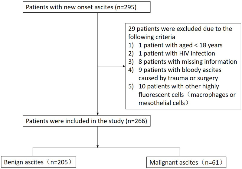
Methods: We collected ascites samples from 266 patients diagnosed with liver cancer. HFC were analyzed using the BF mode of the BC-7500 hematology analyzer, assessing both relative counts (HF-BF%) and absolute counts (HF-BF#). Additionally, biochemical and tumor markers were evaluated in serum and ascites. The diagnostic accuracy of these indicators, both individually and in combination, was assessed using receiver operating characteristic (ROC) curve analysis.
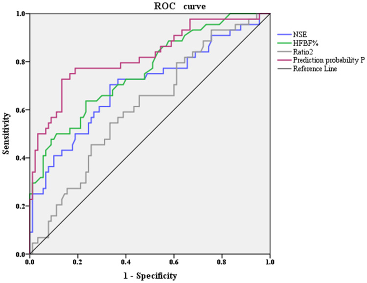
Results: The malignant ascites group exhibited significantly higher levels of HF-BF%, cancer ratio 2 (Ratio2, ascites LDH: ascites ADA Ratio), and neuron-specific enolase (NSE) compared to the benign group, identifying these markers as independent risk factors for malignant ascites in liver cancer patients. Ratio2 demonstrated limited diagnostic value for malignant ascites, with an area under the curve (AUC) of 0.614. In contrast, HF-BF% and NSE showed moderate diagnostic capabilities, with AUCs of 0.760 and 0.700, respectively. The combined assessment of all three indicators yielded a high diagnostic capability, with an AUC of 0.824. The critical values for NSE, HF-BF%, and Ratio2 were 11.42 U/mL, 4.35/100 WBC, and 32.82%, respectively.
Conclusion: The combined evaluation of HF-BF%, Ratio2, and NSE serves as a valuable indicator for predicting the occurrence of metastatic malignant ascites in liver cancer patients.
期刊介绍:
OncoTargets and Therapy is an international, peer-reviewed journal focusing on molecular aspects of cancer research, that is, the molecular diagnosis of and targeted molecular or precision therapy for all types of cancer.
The journal is characterized by the rapid reporting of high-quality original research, basic science, reviews and evaluations, expert opinion and commentary that shed novel insight on a cancer or cancer subtype.
Specific topics covered by the journal include:
-Novel therapeutic targets and innovative agents
-Novel therapeutic regimens for improved benefit and/or decreased side effects
-Early stage clinical trials
Further considerations when submitting to OncoTargets and Therapy:
-Studies containing in vivo animal model data will be considered favorably.
-Tissue microarray analyses will not be considered except in cases where they are supported by comprehensive biological studies involving multiple cell lines.
-Biomarker association studies will be considered only when validated by comprehensive in vitro data and analysis of human tissue samples.
-Studies utilizing publicly available data (e.g. GWAS/TCGA/GEO etc.) should add to the body of knowledge about a specific disease or relevant phenotype and must be validated using the authors’ own data through replication in an independent sample set and functional follow-up.
-Bioinformatics studies must be validated using the authors’ own data through replication in an independent sample set and functional follow-up.
-Single nucleotide polymorphism (SNP) studies will not be considered.
 求助内容:
求助内容: 应助结果提醒方式:


