{"title":"Predictors of diplopia following orbital fractures based on anatomical location, a retrospective cohort study.","authors":"Honglin Chen, Yiqing Bao, Yuxin Chen, Yiqun Li, Peng Wu, Guohua Fan, Ji Zhang","doi":"10.1186/s13005-025-00507-8","DOIUrl":null,"url":null,"abstract":"<p><strong>Purpose: </strong>This study aimed to identify predictors of diplopia following orbital fractures.</p><p><strong>Methods: </strong>We retrospectively analyzed clinical and imaging data from 155 patients who experienced orbital fractures at our center between 2021 and 2023. Orbital fracture sites were classified as C/S/A according to imaging: the lacrimal bone was named as A1, the bony structure behind lamina papyracea as A2 and the lateral wall of the orbit (including the zygomatic bone and the greater wing of sphenoid) was appointed as A3 in the axial view; the orbital floor was divided into three equal parts as S1-S3 in the sagittal view; the frontal process of maxilla was designated as C1, the intermediate central midface between frontal process of maxilla and zygomaticomaxillary suture as C2 and the structure between the zygomaticofrontal suture and the zygomaticomaxillary suture was named as C3. First, we examined clinical characteristics, including age, gender, fracture position, as well as follow-up data on fracture location and diplopia duration. Next, we assessed the correlation between orbital fracture location (C/S/A) and diplopia occurrence. Lastly, we used a multivariable logistic regression model to evaluate predictors associated with the occurrence and location of diplopia in orbital fractures.</p><p><strong>Results: </strong>Among the 155 patients, the mean age was 40.4 ± 14.6 years. Diplopia was the most common ocular symptom after orbital fracture (n = 42, 27.1%). The majority of patients were male (n = 106, 68.4%), with traffic accidents being the leading cause of fractures (n = 107, 69%). Diplopia was observed in 42 patients post-injury. Within the C/S/A classification, only the S region was significantly associated with post-injury diplopia (p = 0.01). Patients with S2, S3, or A1 fractures on preoperative CT had odds ratios (OR) [95% CI] of 2.708 (1.289-5.688), 2.353 (1.141-4.850), and 2.275 (1.068-4.846) for developing diplopia compared to those without these findings. For multiple fracture sites, only sagittal fractures in the S2 + S3 region (p = 0.01) was significantly associated with diplopia. Preoperative A1 fracture was found to increase the likelihood of diplopia by 2.377 times, respectively, according to binary logistic regression analysis.</p><p><strong>Conclusion: </strong>Among the three anatomical views, fractures in the S2, S3, and A1 regions were significantly associated with preoperative diplopia. For patients with multiple fractures, combined S2 and S3 fractures was linked to a higher probability of diplopia. Multivariate analysis indicated that A1 provided the best model for predicting the likelihood of preoperative diplopia.</p>","PeriodicalId":12994,"journal":{"name":"Head & Face Medicine","volume":"21 1","pages":"61"},"PeriodicalIF":2.4000,"publicationDate":"2025-08-18","publicationTypes":"Journal Article","fieldsOfStudy":null,"isOpenAccess":false,"openAccessPdf":"https://www.ncbi.nlm.nih.gov/pmc/articles/PMC12359951/pdf/","citationCount":"0","resultStr":null,"platform":"Semanticscholar","paperid":null,"PeriodicalName":"Head & Face Medicine","FirstCategoryId":"3","ListUrlMain":"https://doi.org/10.1186/s13005-025-00507-8","RegionNum":2,"RegionCategory":"医学","ArticlePicture":[],"TitleCN":null,"AbstractTextCN":null,"PMCID":null,"EPubDate":"","PubModel":"","JCR":"Q2","JCRName":"DENTISTRY, ORAL SURGERY & MEDICINE","Score":null,"Total":0}
引用次数: 0
Abstract
Purpose: This study aimed to identify predictors of diplopia following orbital fractures.
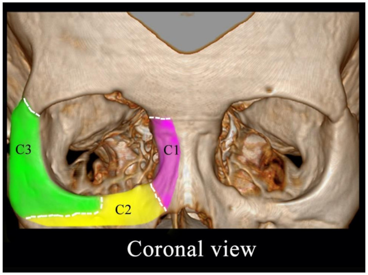
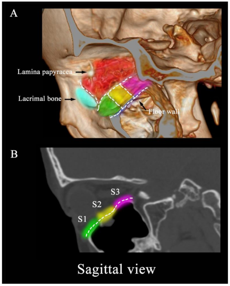
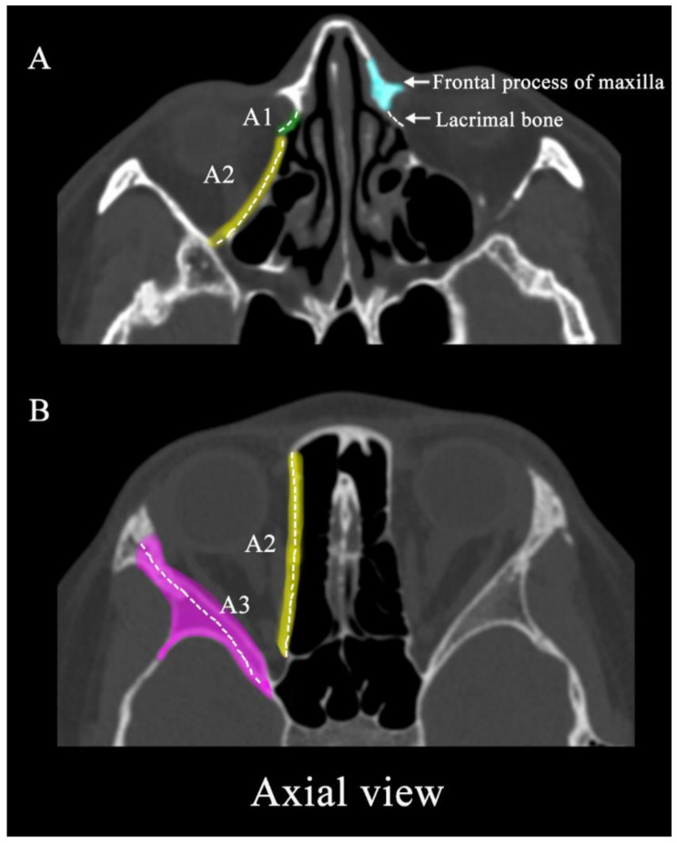
Methods: We retrospectively analyzed clinical and imaging data from 155 patients who experienced orbital fractures at our center between 2021 and 2023. Orbital fracture sites were classified as C/S/A according to imaging: the lacrimal bone was named as A1, the bony structure behind lamina papyracea as A2 and the lateral wall of the orbit (including the zygomatic bone and the greater wing of sphenoid) was appointed as A3 in the axial view; the orbital floor was divided into three equal parts as S1-S3 in the sagittal view; the frontal process of maxilla was designated as C1, the intermediate central midface between frontal process of maxilla and zygomaticomaxillary suture as C2 and the structure between the zygomaticofrontal suture and the zygomaticomaxillary suture was named as C3. First, we examined clinical characteristics, including age, gender, fracture position, as well as follow-up data on fracture location and diplopia duration. Next, we assessed the correlation between orbital fracture location (C/S/A) and diplopia occurrence. Lastly, we used a multivariable logistic regression model to evaluate predictors associated with the occurrence and location of diplopia in orbital fractures.
Results: Among the 155 patients, the mean age was 40.4 ± 14.6 years. Diplopia was the most common ocular symptom after orbital fracture (n = 42, 27.1%). The majority of patients were male (n = 106, 68.4%), with traffic accidents being the leading cause of fractures (n = 107, 69%). Diplopia was observed in 42 patients post-injury. Within the C/S/A classification, only the S region was significantly associated with post-injury diplopia (p = 0.01). Patients with S2, S3, or A1 fractures on preoperative CT had odds ratios (OR) [95% CI] of 2.708 (1.289-5.688), 2.353 (1.141-4.850), and 2.275 (1.068-4.846) for developing diplopia compared to those without these findings. For multiple fracture sites, only sagittal fractures in the S2 + S3 region (p = 0.01) was significantly associated with diplopia. Preoperative A1 fracture was found to increase the likelihood of diplopia by 2.377 times, respectively, according to binary logistic regression analysis.
Conclusion: Among the three anatomical views, fractures in the S2, S3, and A1 regions were significantly associated with preoperative diplopia. For patients with multiple fractures, combined S2 and S3 fractures was linked to a higher probability of diplopia. Multivariate analysis indicated that A1 provided the best model for predicting the likelihood of preoperative diplopia.
期刊介绍:
Head & Face Medicine is a multidisciplinary open access journal that publishes basic and clinical research concerning all aspects of cranial, facial and oral conditions.
The journal covers all aspects of cranial, facial and oral diseases and their management. It has been designed as a multidisciplinary journal for clinicians and researchers involved in the diagnostic and therapeutic aspects of diseases which affect the human head and face. The journal is wide-ranging, covering the development, aetiology, epidemiology and therapy of head and face diseases to the basic science that underlies these diseases. Management of head and face diseases includes all aspects of surgical and non-surgical treatments including psychopharmacological therapies.
 求助内容:
求助内容: 应助结果提醒方式:


