Matthieu Lalevée, Maria Khvesyuk, Andrew Behrens, Philippe Beaudet, Antoine Perrier, Albert DaCosta, François Lintz, Kevin Dibbern, Cesar de Cesar Netto
{"title":"Valgus Deviation of the Intersesamoid Crista in Hallux Valgus and Its Association With the Distal Metatarsal Articular Angle: A Pilot Study.","authors":"Matthieu Lalevée, Maria Khvesyuk, Andrew Behrens, Philippe Beaudet, Antoine Perrier, Albert DaCosta, François Lintz, Kevin Dibbern, Cesar de Cesar Netto","doi":"10.1177/24730114251355501","DOIUrl":null,"url":null,"abstract":"<p><strong>Background: </strong>In hallux valgus (HV), the sesamoid bones embedded in the distal tendon of the flexor hallucis brevis and surrounding the tendon of the flexor hallucis longus are partially guided beneath the head of the first metatarsal (M1) by the intersesamoid crista. The distal metatarsal articular angle (DMAA) assesses the valgus deviation of M1 distal articular surface but is influenced by the pronation of the M1 head relative to the ground. Currently, it is unknown whether the intersesamoid crista itself deviates in valgus in association with the articular surface, and understanding this relationship may have clinical relevance for both the pathogenesis of hallux valgus and its surgical correction.The aim of our study was to compare the angle between the longitudinal axis of the intersesamoid crista and the M1 longitudinal axis in patients with hallux valgus and control subjects and to evaluate its relationship with the DMAA.</p><p><strong>Methods: </strong>A retrospective study was conducted, including 10 HV and 10 matched controls. Weightbearing computed tomography (WBCT) images were automatically segmented with a dedicated software (Disior BoneLogic 2.0) and the angle between the longitudinal axes of the crista and M1 (Crista-M1-angle) as well as the 3d-DMAA (assessing the valgus deviation of the distal articular surface after computerized correction of M1 head pronation relative to the ground) were measured. However, after exclusions for image quality, 9 HV and 8 control feet were analyzed.</p><p><strong>Results: </strong>The mean Crista-M1 angle was deviated in valgus by 14.4 ± 8.7 degrees in 9 HV feet and by 5.5 ± 3.2 degrees in 8 control feet (<i>P</i> = .017). The median 3d-DMAA was deviated in valgus by 9.5 degrees (interquartile range 4.0) in the HV group and by 2.7 degrees (interquartile range 4.5) in controls (<i>P</i> < .001). A positive correlation was observed between Crista-M1 angle and 3d-DMAA (ρ = 0.57; <i>r</i> <sup>2</sup> = 0.328; <i>P</i> = .017).</p><p><strong>Conclusion: </strong>In our pilot study, the longitudinal axis of the intersesamoid crista tended to show greater valgus deviation in HV compared to controls, and this deviation appeared to be correlated with the valgus of M1 distal articular surface. These findings suggest a potential morphologic relationship between crista alignment and distal articular surface orientation. However, clinical implications, such as improved sesamoid tracking, remain speculative.</p><p><strong>Level of evidence: </strong>Level III, retrospective comparative study.</p>","PeriodicalId":12429,"journal":{"name":"Foot & Ankle Orthopaedics","volume":"10 3","pages":"24730114251355501"},"PeriodicalIF":0.0000,"publicationDate":"2025-08-16","publicationTypes":"Journal Article","fieldsOfStudy":null,"isOpenAccess":false,"openAccessPdf":"https://www.ncbi.nlm.nih.gov/pmc/articles/PMC12357992/pdf/","citationCount":"0","resultStr":null,"platform":"Semanticscholar","paperid":null,"PeriodicalName":"Foot & Ankle Orthopaedics","FirstCategoryId":"1085","ListUrlMain":"https://doi.org/10.1177/24730114251355501","RegionNum":0,"RegionCategory":null,"ArticlePicture":[],"TitleCN":null,"AbstractTextCN":null,"PMCID":null,"EPubDate":"2025/7/1 0:00:00","PubModel":"eCollection","JCR":"","JCRName":"","Score":null,"Total":0}
引用次数: 0
Abstract
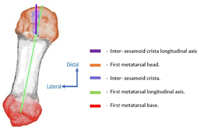
Background: In hallux valgus (HV), the sesamoid bones embedded in the distal tendon of the flexor hallucis brevis and surrounding the tendon of the flexor hallucis longus are partially guided beneath the head of the first metatarsal (M1) by the intersesamoid crista. The distal metatarsal articular angle (DMAA) assesses the valgus deviation of M1 distal articular surface but is influenced by the pronation of the M1 head relative to the ground. Currently, it is unknown whether the intersesamoid crista itself deviates in valgus in association with the articular surface, and understanding this relationship may have clinical relevance for both the pathogenesis of hallux valgus and its surgical correction.The aim of our study was to compare the angle between the longitudinal axis of the intersesamoid crista and the M1 longitudinal axis in patients with hallux valgus and control subjects and to evaluate its relationship with the DMAA.
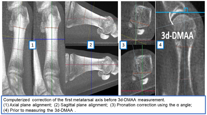
Methods: A retrospective study was conducted, including 10 HV and 10 matched controls. Weightbearing computed tomography (WBCT) images were automatically segmented with a dedicated software (Disior BoneLogic 2.0) and the angle between the longitudinal axes of the crista and M1 (Crista-M1-angle) as well as the 3d-DMAA (assessing the valgus deviation of the distal articular surface after computerized correction of M1 head pronation relative to the ground) were measured. However, after exclusions for image quality, 9 HV and 8 control feet were analyzed.
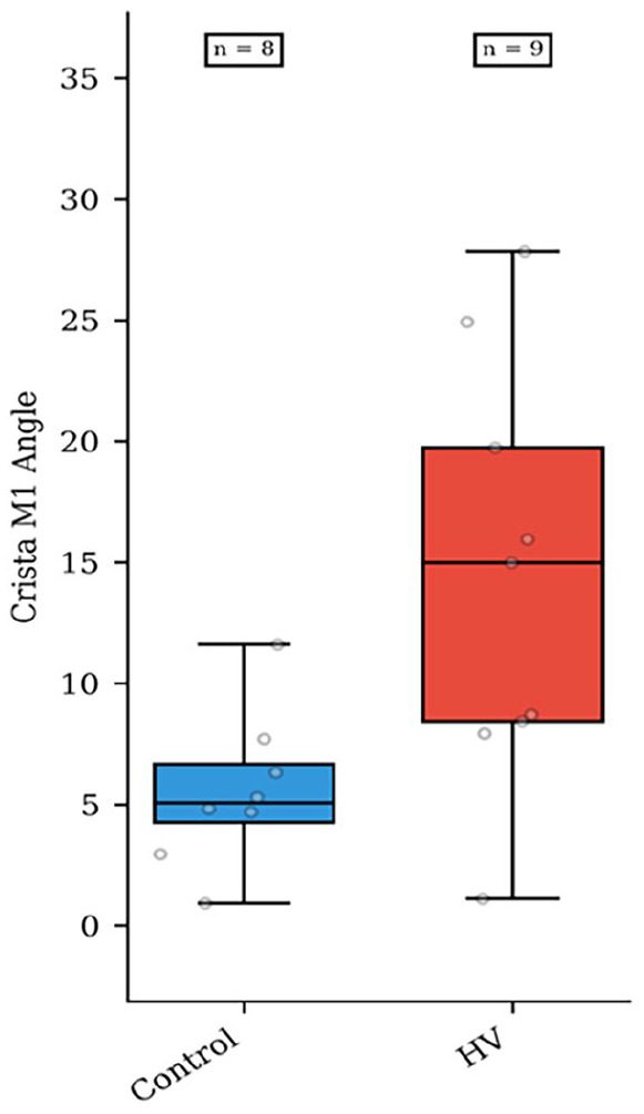
Results: The mean Crista-M1 angle was deviated in valgus by 14.4 ± 8.7 degrees in 9 HV feet and by 5.5 ± 3.2 degrees in 8 control feet (P = .017). The median 3d-DMAA was deviated in valgus by 9.5 degrees (interquartile range 4.0) in the HV group and by 2.7 degrees (interquartile range 4.5) in controls (P < .001). A positive correlation was observed between Crista-M1 angle and 3d-DMAA (ρ = 0.57; r2 = 0.328; P = .017).
Conclusion: In our pilot study, the longitudinal axis of the intersesamoid crista tended to show greater valgus deviation in HV compared to controls, and this deviation appeared to be correlated with the valgus of M1 distal articular surface. These findings suggest a potential morphologic relationship between crista alignment and distal articular surface orientation. However, clinical implications, such as improved sesamoid tracking, remain speculative.
Level of evidence: Level III, retrospective comparative study.

 求助内容:
求助内容: 应助结果提醒方式:


