E. A. Dronova, O. V. Semkin, A. Yu. Shishkin, D. D. Kuklina, A. O. Bogorodskiy, T. S. Kurkin, I. O. Bezruchko, L. M. Vasilenko, Yu. S. Semenov, S. V. Kalenov, A. I. Kuklin, Yu. L. Ryzhykau
{"title":"Wide-Angle X-Ray Scattering Comparative Analysis of Haloarchaeal Purple and Claret Membranes","authors":"E. A. Dronova, O. V. Semkin, A. Yu. Shishkin, D. D. Kuklina, A. O. Bogorodskiy, T. S. Kurkin, I. O. Bezruchko, L. M. Vasilenko, Yu. S. Semenov, S. V. Kalenov, A. I. Kuklin, Yu. L. Ryzhykau","doi":"10.1134/S199074782570028X","DOIUrl":null,"url":null,"abstract":"<p>We present the results of the study of the structural distinctions between the purple membrane (PM) of <i>Halobacterium salinarum</i> and the claret membrane (CM) of <i>Haloquadratum walsbyi</i> using small-angle and wide-angle X-ray scattering. PM and CM are rhodopsin-rich membrane regions. They are crucial for light-driven energy processes in extreme halophilic archaea. While PM structure has been extensively characterized, the structure of CM remains less understood. According to our data, PM exhibits a well-defined hexagonal crystalline lattice with unit cell parameters of <i>a</i> = <i>b</i> ≈ 62 Å, which is consistent with published data. Conversely, CM showed unexpected diffraction patterns, best fitted by rhombohedral lattice with unit cell dimensions <i>a</i> = <i>b</i> = 27.9 Å; γ = 82.9°. But the presence of several unindexed peaks indicates the complexity of the CM sample. These structural differences are attributed to variations in native lipid and pigment compositions, specifically the presence of bacterioruberin in CM, as confirmed by UV-visible spectroscopy. Bacterioruberin, a hallmark of CM, contributes to its red-shifted absorption spectrum, indicative of its organized state within the membrane. However, denaturing 12% polyacrylamide gel electrophoresis (SDS-PAGE) and electron microscopy reveal potential contamination in CM samples, suggesting the need for improved purification protocols. The findings highlight significant structural disparities between PM and CM, with implications for understanding membrane organization. This research underscores the critical role of lipids and pigments in shaping the supramolecular architecture of archaeal membranes and provides a foundation for future studies into their functional adaptations.</p>","PeriodicalId":484,"journal":{"name":"Biochemistry (Moscow), Supplement Series A: Membrane and Cell Biology","volume":"19 3","pages":"331 - 337"},"PeriodicalIF":1.4000,"publicationDate":"2025-08-18","publicationTypes":"Journal Article","fieldsOfStudy":null,"isOpenAccess":false,"openAccessPdf":"","citationCount":"0","resultStr":null,"platform":"Semanticscholar","paperid":null,"PeriodicalName":"Biochemistry (Moscow), Supplement Series A: Membrane and Cell Biology","FirstCategoryId":"2","ListUrlMain":"https://link.springer.com/article/10.1134/S199074782570028X","RegionNum":0,"RegionCategory":null,"ArticlePicture":[],"TitleCN":null,"AbstractTextCN":null,"PMCID":null,"EPubDate":"","PubModel":"","JCR":"Q4","JCRName":"CELL BIOLOGY","Score":null,"Total":0}
引用次数: 0
Abstract
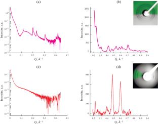
We present the results of the study of the structural distinctions between the purple membrane (PM) of Halobacterium salinarum and the claret membrane (CM) of Haloquadratum walsbyi using small-angle and wide-angle X-ray scattering. PM and CM are rhodopsin-rich membrane regions. They are crucial for light-driven energy processes in extreme halophilic archaea. While PM structure has been extensively characterized, the structure of CM remains less understood. According to our data, PM exhibits a well-defined hexagonal crystalline lattice with unit cell parameters of a = b ≈ 62 Å, which is consistent with published data. Conversely, CM showed unexpected diffraction patterns, best fitted by rhombohedral lattice with unit cell dimensions a = b = 27.9 Å; γ = 82.9°. But the presence of several unindexed peaks indicates the complexity of the CM sample. These structural differences are attributed to variations in native lipid and pigment compositions, specifically the presence of bacterioruberin in CM, as confirmed by UV-visible spectroscopy. Bacterioruberin, a hallmark of CM, contributes to its red-shifted absorption spectrum, indicative of its organized state within the membrane. However, denaturing 12% polyacrylamide gel electrophoresis (SDS-PAGE) and electron microscopy reveal potential contamination in CM samples, suggesting the need for improved purification protocols. The findings highlight significant structural disparities between PM and CM, with implications for understanding membrane organization. This research underscores the critical role of lipids and pigments in shaping the supramolecular architecture of archaeal membranes and provides a foundation for future studies into their functional adaptations.
期刊介绍:
Biochemistry (Moscow), Supplement Series A: Membrane and Cell Biology is an international peer reviewed journal that publishes original articles on physical, chemical, and molecular mechanisms that underlie basic properties of biological membranes and mediate membrane-related cellular functions. The primary topics of the journal are membrane structure, mechanisms of membrane transport, bioenergetics and photobiology, intracellular signaling as well as membrane aspects of cell biology, immunology, and medicine. The journal is multidisciplinary and gives preference to those articles that employ a variety of experimental approaches, basically in biophysics but also in biochemistry, cytology, and molecular biology. The journal publishes articles that strive for unveiling membrane and cellular functions through innovative theoretical models and computer simulations.
 求助内容:
求助内容: 应助结果提醒方式:


