The vascular safe zone for endoscopic third ventriculostomy: a radiomorphometric study
IF 1.8
4区 医学
Q3 CLINICAL NEUROLOGY
引用次数: 0
Abstract
Background and objective
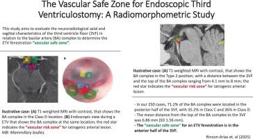
Endoscopic third ventriculostomy (ETV) is a minimally invasive procedure for hydrocephalus management. Variable neuroanatomical features in the floor of the third ventricle (3VF) and the basilar artery (BA) complex contribute to the risk of vascular lesions in the BA complex. This study aims to evaluate the neuroradiological characteristics of the 3VF with the BA complex to determine the ETV fenestration “vascular safe zone”.
Methods
Preoperative neuroimaging studies from a consecutive cohort of adult patients who underwent ETV between January 2016 and March 2025 were retrospectively reviewed. Preoperative MRIs were used to evaluate the neuroradiological axial and sagittal characteristics of the 3VF with the BA complex.
Results
The neuroradiological BA complex position was described. In the sagittal plane, Type II (74 %) was the most common, with mean distances ranging from 4.1 mm to 8 mm. The mean distance from the top of the BA complex to the 3VF was 6.86 mm (SD 1.56 mm). In the axial plane, Classes C and D (71.2 %) were predominant and had varied distances to the 3VF.
Conclusions
Our results suggest that the “vascular safe zone” for an ETV fenestration is primarily located in the anterior half of the 3VF. 71.2 % of our cases showed that the basilar artery (BA) complex was located in the posterior half. Future research should validate the “vascular safe zone” for ETV fenestration with neuroendoscopy intraoperative observations and assess clinical outcomes, including post-operative complications and success rates, to ensure the effectiveness of the proposed fenestration area.
内镜下第三脑室造口术的血管安全区:放射形态学研究
背景与目的内镜下第三脑室造口术(ETV)是治疗脑积水的一种微创手术。第三脑室底(3VF)和基底动脉(BA)复合体多变的神经解剖学特征增加了BA复合体血管病变的风险。本研究旨在评价具有BA复合物的3VF的神经影像学特征,以确定ETV开窗的“血管安全区”。方法回顾性分析2016年1月至2025年3月期间接受ETV手术的连续成年患者的术前神经影像学研究。术前mri评估伴有BA复合体的3VF的轴位和矢状面神经放射学特征。结果描述了神经放射学上BA复合体的位置。在矢状面,II型(74%)最常见,平均距离为4.1 mm至8mm。BA复合物顶部到3VF的平均距离为6.86 mm (SD为1.56 mm)。在轴向面,C类和D类占71.2%,且与3VF的距离不同。结论ETV开窗的“血管安全区”主要位于3VF的前半部分。71.2%的病例显示基底动脉(BA)复合体位于后半部分。未来的研究应通过神经内窥镜术中观察验证ETV开窗的“血管安全区”,并评估临床结果,包括术后并发症和成功率,以确保拟议开窗区域的有效性。
本文章由计算机程序翻译,如有差异,请以英文原文为准。
求助全文
约1分钟内获得全文
求助全文
来源期刊

Journal of Clinical Neuroscience
医学-临床神经学
CiteScore
4.50
自引率
0.00%
发文量
402
审稿时长
40 days
期刊介绍:
This International journal, Journal of Clinical Neuroscience, publishes articles on clinical neurosurgery and neurology and the related neurosciences such as neuro-pathology, neuro-radiology, neuro-ophthalmology and neuro-physiology.
The journal has a broad International perspective, and emphasises the advances occurring in Asia, the Pacific Rim region, Europe and North America. The Journal acts as a focus for publication of major clinical and laboratory research, as well as publishing solicited manuscripts on specific subjects from experts, case reports and other information of interest to clinicians working in the clinical neurosciences.
 求助内容:
求助内容: 应助结果提醒方式:


