A Boronat-López, J Cervera-Ballester, J-C Bernabeu-Mira, M Peñarrocha-Diago, D Peñarrocha-Oltra
{"title":"Changes in the periodontal and tomographic parameters of 36 anterior maxillary teeth one year after periapical surgery with submarginal incision.","authors":"A Boronat-López, J Cervera-Ballester, J-C Bernabeu-Mira, M Peñarrocha-Diago, D Peñarrocha-Oltra","doi":"10.4317/medoral.27157","DOIUrl":null,"url":null,"abstract":"<p><strong>Background: </strong>A study was made of the clinical periodontal changes and buccal cortical bone modifications using cone-beam computed tomography (CBCT) in anterior maxillary teeth with chronic apical periodontitis one year after periapical surgery with submarginal incision.</p><p><strong>Material and methods: </strong>A prospective case series analysis was made of anterior teeth subjected to apical surgery and submarginal incision with a follow-up period of 12 months. Clinical periodontal parameters were recorded, along with tomographic measurements of the buccal cortical bone and volume of the lesion (in mm3) before and one year after surgery. Success was assessed based on the clinical and tomographic data.</p><p><strong>Results: </strong>Thirty-six anterior maxillary teeth from 36 patients with a mean age 43.1 years were enrolled in the study. One year after surgery, mean gingival recession was found to be 0.19 mm with a clinical attachment loss of 0.28 mm. Marginal bone loss was 0.25 mm. The thickness of the buccal cortical bone decreased at all three measurement points, with the greatest decrease being observed at 3 mm from the bone crest (0.58 mm). The distance from the apex to the buccal cortical bone (depth of the apex) decreased 0.59 mm at one year. The clinical parameters (clinical attachment level and probing depth) were not correlated with the tomographic measurements (cementoenamel junction-bone crest distance). The mean lesion volume was 457 mm3 at baseline versus 28.4 mm3 one year after surgery, representing a decrease of 93.8% in 12 months. The success rate at one year postsurgery was 94.4%.</p><p><strong>Conclusions: </strong>One year after apical surgery of anterior maxillary teeth with submarginal incision, only minimal clinical periodontal and tomographic changes are observed, with no clinical relevance. The mean lesion volume decreased 93.8%, and the success rate was 94.4%.</p>","PeriodicalId":49016,"journal":{"name":"Medicina Oral Patologia Oral Y Cirugia Bucal","volume":" ","pages":"e673-e680"},"PeriodicalIF":2.1000,"publicationDate":"2025-09-01","publicationTypes":"Journal Article","fieldsOfStudy":null,"isOpenAccess":false,"openAccessPdf":"https://www.ncbi.nlm.nih.gov/pmc/articles/PMC12395577/pdf/","citationCount":"0","resultStr":null,"platform":"Semanticscholar","paperid":null,"PeriodicalName":"Medicina Oral Patologia Oral Y Cirugia Bucal","FirstCategoryId":"3","ListUrlMain":"https://doi.org/10.4317/medoral.27157","RegionNum":3,"RegionCategory":"医学","ArticlePicture":[],"TitleCN":null,"AbstractTextCN":null,"PMCID":null,"EPubDate":"","PubModel":"","JCR":"Q2","JCRName":"DENTISTRY, ORAL SURGERY & MEDICINE","Score":null,"Total":0}
引用次数: 0
Abstract
Background: A study was made of the clinical periodontal changes and buccal cortical bone modifications using cone-beam computed tomography (CBCT) in anterior maxillary teeth with chronic apical periodontitis one year after periapical surgery with submarginal incision.
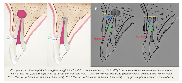
Material and methods: A prospective case series analysis was made of anterior teeth subjected to apical surgery and submarginal incision with a follow-up period of 12 months. Clinical periodontal parameters were recorded, along with tomographic measurements of the buccal cortical bone and volume of the lesion (in mm3) before and one year after surgery. Success was assessed based on the clinical and tomographic data.
Results: Thirty-six anterior maxillary teeth from 36 patients with a mean age 43.1 years were enrolled in the study. One year after surgery, mean gingival recession was found to be 0.19 mm with a clinical attachment loss of 0.28 mm. Marginal bone loss was 0.25 mm. The thickness of the buccal cortical bone decreased at all three measurement points, with the greatest decrease being observed at 3 mm from the bone crest (0.58 mm). The distance from the apex to the buccal cortical bone (depth of the apex) decreased 0.59 mm at one year. The clinical parameters (clinical attachment level and probing depth) were not correlated with the tomographic measurements (cementoenamel junction-bone crest distance). The mean lesion volume was 457 mm3 at baseline versus 28.4 mm3 one year after surgery, representing a decrease of 93.8% in 12 months. The success rate at one year postsurgery was 94.4%.
Conclusions: One year after apical surgery of anterior maxillary teeth with submarginal incision, only minimal clinical periodontal and tomographic changes are observed, with no clinical relevance. The mean lesion volume decreased 93.8%, and the success rate was 94.4%.
期刊介绍:
1. Oral Medicine and Pathology:
Clinicopathological as well as medical or surgical management aspects of
diseases affecting oral mucosa, salivary glands, maxillary bones, as well as
orofacial neurological disorders, and systemic conditions with an impact on
the oral cavity.
2. Oral Surgery:
Surgical management aspects of diseases affecting oral mucosa, salivary glands,
maxillary bones, teeth, implants, oral surgical procedures. Surgical management
of diseases affecting head and neck areas.
3. Medically compromised patients in Dentistry:
Articles discussing medical problems in Odontology will also be included, with
a special focus on the clinico-odontological management of medically compromised patients, and considerations regarding high-risk or disabled patients.
4. Implantology
5. Periodontology

 求助内容:
求助内容: 应助结果提醒方式:


