Shaping Quality in Cardiovascular Magnetic Resonance: A Comparative Study of Segmentation Approaches by Trainees and Experts
IF 2.5
Q2 CARDIAC & CARDIOVASCULAR SYSTEMS
引用次数: 0
Abstract
Background
Cardiovascular magnetic resonance (CMR) is an established cardiovascular imaging (CVI) technique. Deficits in training limit the widespread use of CMR. This study analyzed the influence of CVI experience on segmentation, to define quality standards for teaching and supervision.
Methods
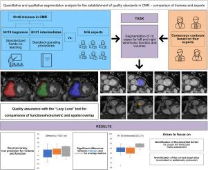
Four CMR experts determined left ventricular (LV) and right ventricular (RV) gold-standard contours in end-systole (ES) and end-diastole (ED), by consensus. After a brief teaching session, readers independently performed segmentations. Readers were classified as beginners (no previous experience in CVI), intermediates (previous experience in CVI, but not in CMR), or experts (extensive experience in CVI including CMR). Results were compared, and the cause of deviation was analyzed, using metrics such as the Dice similarity coefficient (DSC).
Results
A total of 46 readers (19 beginners, 21 intermediates, 6 experts) performed image analysis. Using the DSC, we found significant differences in endocardial LV ED contours (median [interquartile range]: beginners, 92.9% [91.9%-93.5%]); intermediates, 93.5% (93.0%-94.1%); experts, 93.9% (93.1%-94.3%); P = 0.043) and in myocardial contours (beginners, 79.0% (75.0%-80.9%); intermediates, 80.9% (78.0%-82.4%); experts, 85.0% (79.8%-86.5%); p = 0.001). Experts had higher DSC scores for the right ventricle (ES: beginners, 83.8% (81.3%-85.8%); intermediates, 81.7% (79.6%-85.6%); experts, 89.0% (86.6%-89.8%); P = 0.003; ED: beginners, 89.2% (88.1%-90.3%); intermediates, 88.6% (87.9%-89.2%); experts, 91.6% (89.8%-93.3%); P = 0.002). The disagreements were not traceable in absolute volume and function (P for all > 0.2). Sources of disagreement were related mainly to handling of basal slices.
Conclusions
After a brief standardized teaching session, beginners and intermediates performed chamber quantification consistent with that of experts. Differences, especially in LV mass and RV segmentations, warrant continuous training, ideally accompanied by automatic methods for quality assurance.
心血管磁共振整形质量:学员和专家分割方法的比较研究
心血管磁共振(CMR)是一种成熟的心血管成像(CVI)技术。培训方面的缺陷限制了CMR的广泛应用。本研究分析CVI经验对分割的影响,以厘定教学与督导的品质标准。方法4位CMR专家一致确定收缩期末(ES)和舒张期末(ED)左心室(LV)和右心室(RV)的金标准轮廓。在一个简短的教学环节后,读者独立地进行分段。读者被分为初学者(以前没有CVI经验)、中级(以前有CVI经验,但没有CMR经验)或专家(在CVI包括CMR方面有丰富经验)。使用Dice相似系数(DSC)等指标对结果进行比较,并分析偏差的原因。结果共46名读者进行了图像分析,其中初学者19人,中级21人,专家6人。使用DSC,我们发现心内膜内LV ED轮廓有显著差异(中位数[四分位数范围]:初学者,92.9% [91.9%-93.5%]);中间体93.5% (93.0%-94.1%);专家占93.9% (93.1% ~ 94.3%);P = 0.043)和心肌轮廓(初学者79.0% (75.0% ~ 80.9%);中间体占80.9% (78.0% ~ 82.4%);专家占85.0% (79.8% ~ 86.5%);P = 0.001)。专家右心室DSC得分较高(ES:初学者,83.8% (81.3% ~ 85.8%);中间体占81.7% (79.6%-85.6%);专家占89.0% (86.6% ~ 89.8%);P = 0.003;ED:初学者89.2% (88.1%-90.3%);中间体占88.6% (87.9%-89.2%);专家占91.6% (89.8% ~ 93.3%);P = 0.002)。分歧在绝对体积和功能上是不可追溯的(P for all >;0.2)。分歧的来源主要与处理基底切片有关。结论经过简短的标准化教学后,初学者和中级学员的室量化操作与专家一致。差异,特别是左室质量和右室分割的差异,需要持续的训练,最好是伴随着质量保证的自动方法。
本文章由计算机程序翻译,如有差异,请以英文原文为准。
求助全文
约1分钟内获得全文
求助全文
来源期刊

CJC Open
Medicine-Cardiology and Cardiovascular Medicine
CiteScore
3.30
自引率
0.00%
发文量
143
审稿时长
60 days
 求助内容:
求助内容: 应助结果提醒方式:


