Manlio Fabio Lara-Duck, Juan Rosales-Martínez, Netzahualcoyotl Mayek-Pérez
{"title":"[Wolff-Parkinson-White syndrome due to cocaine use: a case report].","authors":"Manlio Fabio Lara-Duck, Juan Rosales-Martínez, Netzahualcoyotl Mayek-Pérez","doi":"10.5281/zenodo.16748296","DOIUrl":null,"url":null,"abstract":"<p><strong>Background: </strong>Wolff-Parkinson-White syndrome (WPWS) causes an accessory pathway between the atria and ventricles, in parallel with the atrioventricular node and the bundle of His; it causes a \"short circuit\" that deregulates the physiological pacing and causes tachycardia. A case of WPWS is described in a patient with cocaine, alcohol, and tobacco consumption.</p><p><strong>Clinical case: </strong>A 34-year-old man with a regular history of cocaine, alcohol, and tobacco use for 14 years presented with paroxysmal palpitations, chest pain, and shortness of breath. ECG 1 revealed supraventricular tachycardia with delta waves and a short P-R interval; ECG 2 revealed reversal of the tachycardia without delta waves and inverted T waves in lead 1, aVR, and aVL, and a short P-R interval; ECG 3 revealed sinus rhythm, 90 bpm, and inverted T waves in lead 3, aVR, and v1. TTE showed a normal left ventricle; a normal LVEF, greater than 50% (Simpson's method); and no stenosis or regurgitation in the mitral, tricuspid, and mitral valves. 24-hour Holter monitoring revealed non-sustained paroxysmal supraventricular tachycardia, with a narrow QRS complex, a short P-R, interval of 122 bpm, and T wave inversion during paroxysm. Reversal of paroxysm maintained a short P-R and normal T waves.</p><p><strong>Conclusions: </strong>Upon reversal of the patient's supraventricular tachycardia paroxysms, his T waves corrected (positive T waves). The inverted T waves were due to WPWS, triggered by cocaine, alcohol, and tobacco use, and possibly related to myocardial ischemic involvement.</p>","PeriodicalId":94200,"journal":{"name":"Revista medica del Instituto Mexicano del Seguro Social","volume":"63 5","pages":"e6679"},"PeriodicalIF":0.0000,"publicationDate":"2025-08-14","publicationTypes":"Journal Article","fieldsOfStudy":null,"isOpenAccess":false,"openAccessPdf":"https://www.ncbi.nlm.nih.gov/pmc/articles/PMC12380318/pdf/","citationCount":"0","resultStr":null,"platform":"Semanticscholar","paperid":null,"PeriodicalName":"Revista medica del Instituto Mexicano del Seguro Social","FirstCategoryId":"1085","ListUrlMain":"https://doi.org/10.5281/zenodo.16748296","RegionNum":0,"RegionCategory":null,"ArticlePicture":[],"TitleCN":null,"AbstractTextCN":null,"PMCID":null,"EPubDate":"","PubModel":"","JCR":"","JCRName":"","Score":null,"Total":0}
引用次数: 0
Abstract
Background: Wolff-Parkinson-White syndrome (WPWS) causes an accessory pathway between the atria and ventricles, in parallel with the atrioventricular node and the bundle of His; it causes a "short circuit" that deregulates the physiological pacing and causes tachycardia. A case of WPWS is described in a patient with cocaine, alcohol, and tobacco consumption.
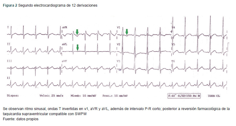
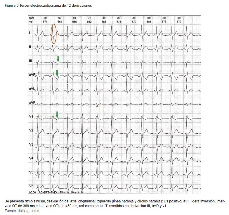
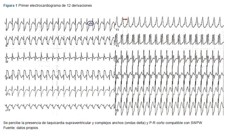
Clinical case: A 34-year-old man with a regular history of cocaine, alcohol, and tobacco use for 14 years presented with paroxysmal palpitations, chest pain, and shortness of breath. ECG 1 revealed supraventricular tachycardia with delta waves and a short P-R interval; ECG 2 revealed reversal of the tachycardia without delta waves and inverted T waves in lead 1, aVR, and aVL, and a short P-R interval; ECG 3 revealed sinus rhythm, 90 bpm, and inverted T waves in lead 3, aVR, and v1. TTE showed a normal left ventricle; a normal LVEF, greater than 50% (Simpson's method); and no stenosis or regurgitation in the mitral, tricuspid, and mitral valves. 24-hour Holter monitoring revealed non-sustained paroxysmal supraventricular tachycardia, with a narrow QRS complex, a short P-R, interval of 122 bpm, and T wave inversion during paroxysm. Reversal of paroxysm maintained a short P-R and normal T waves.
Conclusions: Upon reversal of the patient's supraventricular tachycardia paroxysms, his T waves corrected (positive T waves). The inverted T waves were due to WPWS, triggered by cocaine, alcohol, and tobacco use, and possibly related to myocardial ischemic involvement.

 求助内容:
求助内容: 应助结果提醒方式:


