Zhenshan Gao, Qiang Ma, Lifang Zhu, Dejin Kong, Mingyan Ji, Caipeng Gao, Bo Tang, Zhiqiang Dong
{"title":"Abnormal brain morphology and morphological brain network in adults with acute mild traumatic brain injury.","authors":"Zhenshan Gao, Qiang Ma, Lifang Zhu, Dejin Kong, Mingyan Ji, Caipeng Gao, Bo Tang, Zhiqiang Dong","doi":"10.1097/WNR.0000000000002186","DOIUrl":null,"url":null,"abstract":"<p><strong>Background: </strong>Mild traumatic brain injury (mTBI) commonly has long-term cognitive and functional consequences; however, it is not clear whether these adverse outcomes begin in the acute phase of mTBI and are associated with changes in brain morphology and function.</p><p><strong>Methods: </strong>The current study used T1-weighted MRI to determine whether cortical thickness, gray matter volume (GMV), and morphological brain networks were altered in patients with mTBI within 7 days of injury, and to examine whether these changes were associated with postacute cognitive and emotional abnormalities. Adults aged 18-56 years with mTBI ( n = 43) and healthy controls ( n = 37) completed the cognitive, emotional evaluation, and MRI examination, during which patients with mTBI completed symptom reports. Cortical thickness and GMV were estimated using Computational Anatomy Toolbox 12. On this basis, a gray matter covariance network was constructed based on the cortical thickness.</p><p><strong>Results: </strong>After false discovery rate (FDR) correction, groups differed significantly on the left parahippocampal gyrus and left orbital part of the superior frontal gyrus GMV (mTBI > controls), but no cortical thickness. The network topological properties were also changed in the acute stage of mTBI. The GMV abnormality was related to postacute cognitive and emotional changes in the mTBI group.</p><p><strong>Conclusion: </strong>The results emphasize that adverse outcomes begin in the acute phase and that the left parahippocampal gyrus and left orbital part of the superior frontal gyrus and related brain network abnormalities may be potential neuroimaging biomarkers explaining acute cognitive and depressive symptoms.</p>","PeriodicalId":19213,"journal":{"name":"Neuroreport","volume":" ","pages":"777-785"},"PeriodicalIF":1.7000,"publicationDate":"2025-10-01","publicationTypes":"Journal Article","fieldsOfStudy":null,"isOpenAccess":false,"openAccessPdf":"https://www.ncbi.nlm.nih.gov/pmc/articles/PMC12393062/pdf/","citationCount":"0","resultStr":null,"platform":"Semanticscholar","paperid":null,"PeriodicalName":"Neuroreport","FirstCategoryId":"3","ListUrlMain":"https://doi.org/10.1097/WNR.0000000000002186","RegionNum":4,"RegionCategory":"医学","ArticlePicture":[],"TitleCN":null,"AbstractTextCN":null,"PMCID":null,"EPubDate":"2025/7/21 0:00:00","PubModel":"Epub","JCR":"Q4","JCRName":"NEUROSCIENCES","Score":null,"Total":0}
引用次数: 0
Abstract
Background: Mild traumatic brain injury (mTBI) commonly has long-term cognitive and functional consequences; however, it is not clear whether these adverse outcomes begin in the acute phase of mTBI and are associated with changes in brain morphology and function.
Methods: The current study used T1-weighted MRI to determine whether cortical thickness, gray matter volume (GMV), and morphological brain networks were altered in patients with mTBI within 7 days of injury, and to examine whether these changes were associated with postacute cognitive and emotional abnormalities. Adults aged 18-56 years with mTBI ( n = 43) and healthy controls ( n = 37) completed the cognitive, emotional evaluation, and MRI examination, during which patients with mTBI completed symptom reports. Cortical thickness and GMV were estimated using Computational Anatomy Toolbox 12. On this basis, a gray matter covariance network was constructed based on the cortical thickness.
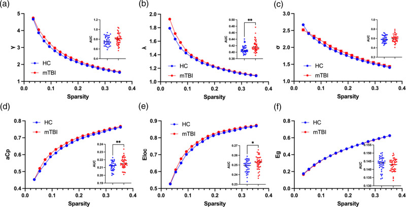
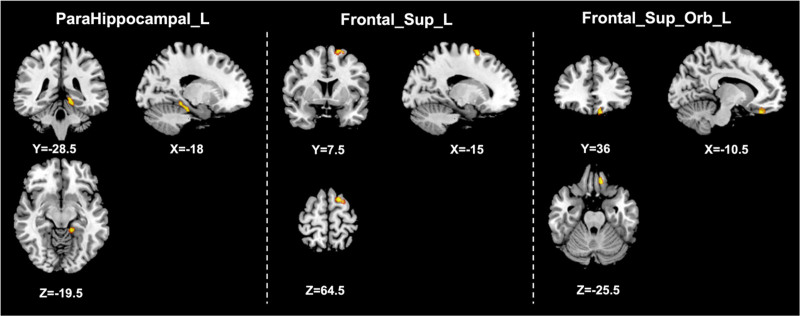
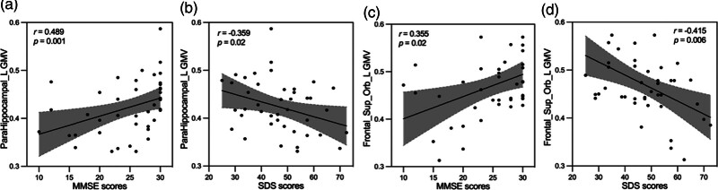
Results: After false discovery rate (FDR) correction, groups differed significantly on the left parahippocampal gyrus and left orbital part of the superior frontal gyrus GMV (mTBI > controls), but no cortical thickness. The network topological properties were also changed in the acute stage of mTBI. The GMV abnormality was related to postacute cognitive and emotional changes in the mTBI group.
Conclusion: The results emphasize that adverse outcomes begin in the acute phase and that the left parahippocampal gyrus and left orbital part of the superior frontal gyrus and related brain network abnormalities may be potential neuroimaging biomarkers explaining acute cognitive and depressive symptoms.
期刊介绍:
NeuroReport is a channel for rapid communication of new findings in neuroscience. It is a forum for the publication of short but complete reports of important studies that require very fast publication. Papers are accepted on the basis of the novelty of their finding, on their significance for neuroscience and on a clear need for rapid publication. Preliminary communications are not suitable for the Journal. Submitted articles undergo a preliminary review by the editor. Some articles may be returned to authors without further consideration. Those being considered for publication will undergo further assessment and peer-review by the editors and those invited to do so from a reviewer pool.
The core interest of the Journal is on studies that cast light on how the brain (and the whole of the nervous system) works.
We aim to give authors a decision on their submission within 2-5 weeks, and all accepted articles appear in the next issue to press.
 求助内容:
求助内容: 应助结果提醒方式:


