Ayşe Dilaver Akar, Nami Yeyin, Sinem Akyol, Özge Demir, Eylem Gülce Çoker, Mustafa Demir
{"title":"Investigation of the Partial Volume Effect in Pre-Dosimetry of Liver Tumors for <sup>90</sup>Y Radioembolization: A Phantom Study.","authors":"Ayşe Dilaver Akar, Nami Yeyin, Sinem Akyol, Özge Demir, Eylem Gülce Çoker, Mustafa Demir","doi":"10.4274/mirt.galenos.2025.77200","DOIUrl":null,"url":null,"abstract":"<p><strong>Objectives: </strong>Yttrium-90 (<sup>90</sup>Y) radioembolization has become increasingly important in the treatment of liver tumors. This study aims to experimentally determine the extent to which small liver tumors are affected by the partial volume effect (PVE) in single photon emission computed tomography/computed tomography (SPECT/CT) scintigraphy using technetium-99m-macroaggregated albumin (Tc-99m-MAA), and to investigate the impact of PVE on tumor dosimetry and image quality.</p><p><strong>Methods: </strong>In this experimental study, a custom-designed liver phantom containing four tumor mimics with diameters of 1 cm, 2 cm, 3 cm, and 5 cm was used. The tumor and liver parenchyma volumes were filled with Tc-99m at a ratio of 4.86: 1. The phantom was imaged in a water tank using SPECT/CT according to standard clinical protocols. Volumetric regions of interest were drawn for each lesion and tumor volumes, contrast values (C), contrast to noise ratios (CNR), and absorbed tumor doses were calculated from the counts obtained. Since this study does not involve live subjects and was conducted solely on a phantom model, ethical approval, informed consent, and consent forms are not required for this study.</p><p><strong>Results: </strong>Tumor diameters measured on SPECT/CT images matched those obtained from both CT images and the actual dimensions. The contrast values calculated from the SPECT/CT images for lesions with diameters of 2 cm and 5 cm were 2.03 and 3.89, respectively. Similarly, the corresponding CNR values were 8.64 and 21.07. Tumor-to-normal tissue ratios were 2.03 and 3.89 for the 2 cm and 5 cm lesions, respectively. For the 2 cm lesion, the actual and SPECT/CT-derived absorbed doses were 15.3 Gy and 7.87 Gy, respectively. For the 5 cm lesion, these values were 15.4 Gy and 13.38 Gy, respectively. The absorbed tumor doses significantly decreased as tumor diameter decreased due to the influence of PVE.</p><p><strong>Conclusion: </strong>Tumors smaller than 2 cm in diameter were markedly affected by the PVE. Considering the influence of PVE, or applying appropriate corrections in dosimetric calculations, is of critical importance for improving the accuracy of dosimetry results.</p>","PeriodicalId":44681,"journal":{"name":"Molecular Imaging and Radionuclide Therapy","volume":" ","pages":"180-187"},"PeriodicalIF":1.1000,"publicationDate":"2025-10-08","publicationTypes":"Journal Article","fieldsOfStudy":null,"isOpenAccess":false,"openAccessPdf":"https://www.ncbi.nlm.nih.gov/pmc/articles/PMC12505186/pdf/","citationCount":"0","resultStr":null,"platform":"Semanticscholar","paperid":null,"PeriodicalName":"Molecular Imaging and Radionuclide Therapy","FirstCategoryId":"1085","ListUrlMain":"https://doi.org/10.4274/mirt.galenos.2025.77200","RegionNum":0,"RegionCategory":null,"ArticlePicture":[],"TitleCN":null,"AbstractTextCN":null,"PMCID":null,"EPubDate":"2025/8/13 0:00:00","PubModel":"Epub","JCR":"Q4","JCRName":"RADIOLOGY, NUCLEAR MEDICINE & MEDICAL IMAGING","Score":null,"Total":0}
引用次数: 0
Abstract
Objectives: Yttrium-90 (90Y) radioembolization has become increasingly important in the treatment of liver tumors. This study aims to experimentally determine the extent to which small liver tumors are affected by the partial volume effect (PVE) in single photon emission computed tomography/computed tomography (SPECT/CT) scintigraphy using technetium-99m-macroaggregated albumin (Tc-99m-MAA), and to investigate the impact of PVE on tumor dosimetry and image quality.
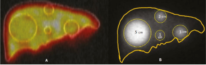
Methods: In this experimental study, a custom-designed liver phantom containing four tumor mimics with diameters of 1 cm, 2 cm, 3 cm, and 5 cm was used. The tumor and liver parenchyma volumes were filled with Tc-99m at a ratio of 4.86: 1. The phantom was imaged in a water tank using SPECT/CT according to standard clinical protocols. Volumetric regions of interest were drawn for each lesion and tumor volumes, contrast values (C), contrast to noise ratios (CNR), and absorbed tumor doses were calculated from the counts obtained. Since this study does not involve live subjects and was conducted solely on a phantom model, ethical approval, informed consent, and consent forms are not required for this study.
Results: Tumor diameters measured on SPECT/CT images matched those obtained from both CT images and the actual dimensions. The contrast values calculated from the SPECT/CT images for lesions with diameters of 2 cm and 5 cm were 2.03 and 3.89, respectively. Similarly, the corresponding CNR values were 8.64 and 21.07. Tumor-to-normal tissue ratios were 2.03 and 3.89 for the 2 cm and 5 cm lesions, respectively. For the 2 cm lesion, the actual and SPECT/CT-derived absorbed doses were 15.3 Gy and 7.87 Gy, respectively. For the 5 cm lesion, these values were 15.4 Gy and 13.38 Gy, respectively. The absorbed tumor doses significantly decreased as tumor diameter decreased due to the influence of PVE.
Conclusion: Tumors smaller than 2 cm in diameter were markedly affected by the PVE. Considering the influence of PVE, or applying appropriate corrections in dosimetric calculations, is of critical importance for improving the accuracy of dosimetry results.
期刊介绍:
Molecular Imaging and Radionuclide Therapy (Mol Imaging Radionucl Ther, MIRT) is publishes original research articles, invited reviews, editorials, short communications, letters, consensus statements, guidelines and case reports with a literature review on the topic, in the field of molecular imaging, multimodality imaging, nuclear medicine, radionuclide therapy, radiopharmacy, medical physics, dosimetry and radiobiology.


 求助内容:
求助内容: 应助结果提醒方式:


