Li Wang, Yingqian Tan, Fang Li, Yue Liang, Guangui Chen, Amy Michelle Huang, Lina Li, Junming Wang, Zhiping Liu
{"title":"Unilateral Painless Visual Loss in Sphenoid Mucoceles with Optic Neuropathy.","authors":"Li Wang, Yingqian Tan, Fang Li, Yue Liang, Guangui Chen, Amy Michelle Huang, Lina Li, Junming Wang, Zhiping Liu","doi":"10.1159/000546758","DOIUrl":null,"url":null,"abstract":"<p><strong>Introduction: </strong>Mucoceles are benign, slow-growing cystic formations located within the paranasal sinuses, caused by complete ostial obstruction and accumulation of mucous secretions. Here, we report a case of a patient who initially presented with unilateral painless visual impairment and was ultimately diagnosed with bilateral sphenoid sinus mucoceles (SSMs) after two hospitalizations.</p><p><strong>Case presentation: </strong>A 67-year-old woman presented with a 7-day history of decreased vision in her left eye. She denied any orbital pain, headache, or restricted eye movement and was diagnosed with retrobulbar ischemic optic neuropathy on the first admission. After drug treatment, the visual acuity of patient improved significantly, but 3 months after discharge, the patient was hospitalized again due to recurrent vision loss accompanied by left orbital pain and left temporal pulsatile headaches. After multiple imaging examinations, the patient was ultimately diagnosed with SSMs and her visual acuity was restored after surgical treatment.</p><p><strong>Conclusions: </strong>The majority of SSMs are associated with ocular symptoms, with only a minority presenting solely with unilateral or bilateral vision loss, as exemplified in this case. Therefore, understanding the clinical features of visual disturbances secondary to SSMs is crucial to aiding more prompt diagnosis and treatment.</p>","PeriodicalId":9635,"journal":{"name":"Case Reports in Ophthalmology","volume":"16 1","pages":"559-566"},"PeriodicalIF":0.6000,"publicationDate":"2025-07-07","publicationTypes":"Journal Article","fieldsOfStudy":null,"isOpenAccess":false,"openAccessPdf":"https://www.ncbi.nlm.nih.gov/pmc/articles/PMC12342703/pdf/","citationCount":"0","resultStr":null,"platform":"Semanticscholar","paperid":null,"PeriodicalName":"Case Reports in Ophthalmology","FirstCategoryId":"1085","ListUrlMain":"https://doi.org/10.1159/000546758","RegionNum":0,"RegionCategory":null,"ArticlePicture":[],"TitleCN":null,"AbstractTextCN":null,"PMCID":null,"EPubDate":"2025/1/1 0:00:00","PubModel":"eCollection","JCR":"Q4","JCRName":"OPHTHALMOLOGY","Score":null,"Total":0}
引用次数: 0
Abstract
Introduction: Mucoceles are benign, slow-growing cystic formations located within the paranasal sinuses, caused by complete ostial obstruction and accumulation of mucous secretions. Here, we report a case of a patient who initially presented with unilateral painless visual impairment and was ultimately diagnosed with bilateral sphenoid sinus mucoceles (SSMs) after two hospitalizations.
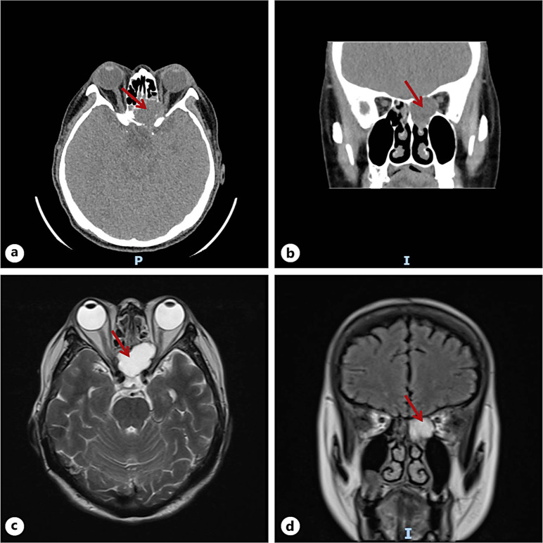
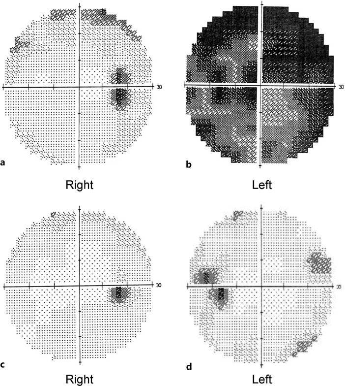
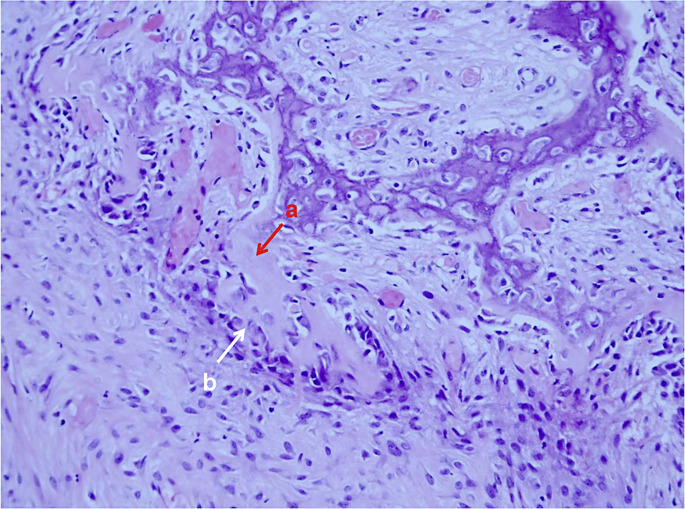
Case presentation: A 67-year-old woman presented with a 7-day history of decreased vision in her left eye. She denied any orbital pain, headache, or restricted eye movement and was diagnosed with retrobulbar ischemic optic neuropathy on the first admission. After drug treatment, the visual acuity of patient improved significantly, but 3 months after discharge, the patient was hospitalized again due to recurrent vision loss accompanied by left orbital pain and left temporal pulsatile headaches. After multiple imaging examinations, the patient was ultimately diagnosed with SSMs and her visual acuity was restored after surgical treatment.
Conclusions: The majority of SSMs are associated with ocular symptoms, with only a minority presenting solely with unilateral or bilateral vision loss, as exemplified in this case. Therefore, understanding the clinical features of visual disturbances secondary to SSMs is crucial to aiding more prompt diagnosis and treatment.
期刊介绍:
This peer-reviewed online-only journal publishes original case reports covering the entire spectrum of ophthalmology, including prevention, diagnosis, treatment, toxicities of therapy, supportive care, quality-of-life, and survivorship issues. The submission of negative results is strongly encouraged. The journal will also accept case reports dealing with the use of novel technologies, both in the arena of diagnosis and treatment. Supplementary material is welcomed. The intent of the journal is to provide clinicians and researchers with a tool to disseminate their personal experiences to a wider public as well as to review interesting cases encountered by colleagues all over the world. Universally used terms can be searched across the entire growing collection of case reports, further facilitating the retrieval of specific information. Following the open access principle, the entire contents can be retrieved at no charge, guaranteeing easy access to this valuable source of anecdotal information at all times.

 求助内容:
求助内容: 应助结果提醒方式:


