Predicting preoperative lymph node metastasis in patients with high-grade serous ovarian cancer by using intratumoral and peritumoral radiomics: a retrospective cohort study.
IF 3.2 3区 医学Q2 RADIOLOGY, NUCLEAR MEDICINE & MEDICAL IMAGING
Silin Nie, Yumin Jiang, Huixiang Ji, Xiaohui Liu, Lanxing Lyu, Chun Wang, Yuping Shan, Aiping Chen
{"title":"Predicting preoperative lymph node metastasis in patients with high-grade serous ovarian cancer by using intratumoral and peritumoral radiomics: a retrospective cohort study.","authors":"Silin Nie, Yumin Jiang, Huixiang Ji, Xiaohui Liu, Lanxing Lyu, Chun Wang, Yuping Shan, Aiping Chen","doi":"10.1186/s12880-025-01868-x","DOIUrl":null,"url":null,"abstract":"<p><strong>Background: </strong>Ovarian cancer (OC) carries the worst prognosis among gynecologic cancers, with high-grade serous ovarian cancer (HGSOC) as its most common subtype. Cytoreductive surgery (tumor resection) is the cornerstone of OC treatment. However, controversy remains regarding whether lymphadenectomy should be performed during surgery; more than 30% of patients with OC undergo unnecessary lymphadenectomy, increasing surgical risks and prolonging postoperative recovery. By analyzing multidimensional imaging features, such as tumor morphology, texture, and density, radiomics can accurately quantify the biological characteristics of tumors. However, its application in OC needs to be explored further. This study aimed to explore radiomics' role in predicting lymph node metastasis risk in HGSOC.</p><p><strong>Methods: </strong>This retrospective cohort analysis involved 273 participants from Qingdao University Affiliated Hospital and Rizhao People's Hospital, and they were categorized into the training, testing, and external validation groups. Imaging characteristics were derived from the tumor region of interest and its surrounding areas (1-5 mm), and radiomics scores were calculated for each region. This approach was employed for assessing the diagnostic performance of different regions and identify the optimal one. We constructed a risk prediction model that integrated imaging features of the optimal region with independent clinical risk factors.</p><p><strong>Results: </strong>The radiomic features of the tumor and its surrounding 3-mm extension region yielded area under the curve (AUC) values of 0.957 and 0.793 in the training and testing sets, respectively. After integrating the radiomic features of the tumor and its surrounding 3-mm extension region with clinical features, the AUC values in the training set, testing set, and external validation set were 0.971, 0.811, and 0.869, respectively, demonstrating strong predictive ability.</p><p><strong>Conclusions: </strong>This study developed a model to assess lymph node metastasis likelihood in HGSOC patients. In the test and external validation cohorts, the model demonstrated excellent predictive performance. We believe the model can assist clinicians in identifying patients who are suitable for lymph node resection, thereby optimizing treatment decisions.</p><p><strong>Clinical trial number: </strong>Not applicable.</p>","PeriodicalId":9020,"journal":{"name":"BMC Medical Imaging","volume":"25 1","pages":"323"},"PeriodicalIF":3.2000,"publicationDate":"2025-08-12","publicationTypes":"Journal Article","fieldsOfStudy":null,"isOpenAccess":false,"openAccessPdf":"https://www.ncbi.nlm.nih.gov/pmc/articles/PMC12341290/pdf/","citationCount":"0","resultStr":null,"platform":"Semanticscholar","paperid":null,"PeriodicalName":"BMC Medical Imaging","FirstCategoryId":"3","ListUrlMain":"https://doi.org/10.1186/s12880-025-01868-x","RegionNum":3,"RegionCategory":"医学","ArticlePicture":[],"TitleCN":null,"AbstractTextCN":null,"PMCID":null,"EPubDate":"","PubModel":"","JCR":"Q2","JCRName":"RADIOLOGY, NUCLEAR MEDICINE & MEDICAL IMAGING","Score":null,"Total":0}
引用次数: 0
Abstract
Background: Ovarian cancer (OC) carries the worst prognosis among gynecologic cancers, with high-grade serous ovarian cancer (HGSOC) as its most common subtype. Cytoreductive surgery (tumor resection) is the cornerstone of OC treatment. However, controversy remains regarding whether lymphadenectomy should be performed during surgery; more than 30% of patients with OC undergo unnecessary lymphadenectomy, increasing surgical risks and prolonging postoperative recovery. By analyzing multidimensional imaging features, such as tumor morphology, texture, and density, radiomics can accurately quantify the biological characteristics of tumors. However, its application in OC needs to be explored further. This study aimed to explore radiomics' role in predicting lymph node metastasis risk in HGSOC.
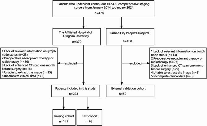
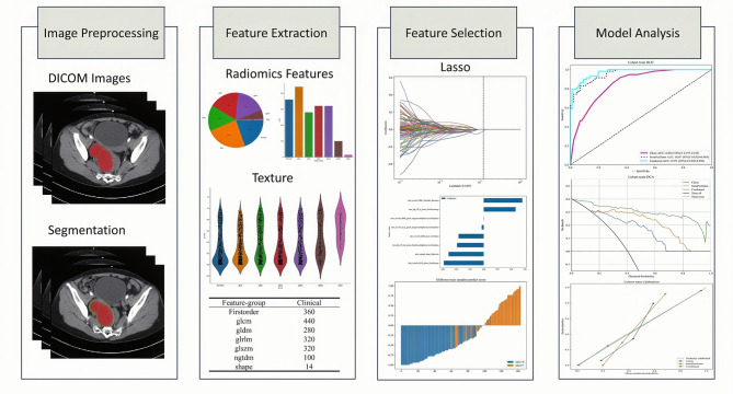
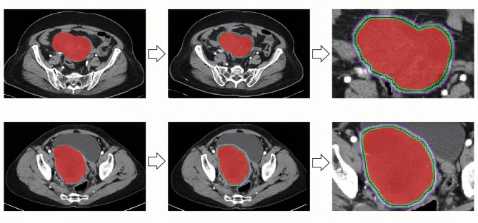
Methods: This retrospective cohort analysis involved 273 participants from Qingdao University Affiliated Hospital and Rizhao People's Hospital, and they were categorized into the training, testing, and external validation groups. Imaging characteristics were derived from the tumor region of interest and its surrounding areas (1-5 mm), and radiomics scores were calculated for each region. This approach was employed for assessing the diagnostic performance of different regions and identify the optimal one. We constructed a risk prediction model that integrated imaging features of the optimal region with independent clinical risk factors.
Results: The radiomic features of the tumor and its surrounding 3-mm extension region yielded area under the curve (AUC) values of 0.957 and 0.793 in the training and testing sets, respectively. After integrating the radiomic features of the tumor and its surrounding 3-mm extension region with clinical features, the AUC values in the training set, testing set, and external validation set were 0.971, 0.811, and 0.869, respectively, demonstrating strong predictive ability.
Conclusions: This study developed a model to assess lymph node metastasis likelihood in HGSOC patients. In the test and external validation cohorts, the model demonstrated excellent predictive performance. We believe the model can assist clinicians in identifying patients who are suitable for lymph node resection, thereby optimizing treatment decisions.
期刊介绍:
BMC Medical Imaging is an open access journal publishing original peer-reviewed research articles in the development, evaluation, and use of imaging techniques and image processing tools to diagnose and manage disease.

 求助内容:
求助内容: 应助结果提醒方式:


