Shilpa Levingston, Shivaprasad, Devika Shetty, Aneesa Ayoob, Shruthi M
{"title":"Assessment of the submandibular fossa depth and diameter of the mandibular canal via cone beam computed tomography: a comparative study.","authors":"Shilpa Levingston, Shivaprasad, Devika Shetty, Aneesa Ayoob, Shruthi M","doi":"10.1186/s40902-025-00473-w","DOIUrl":null,"url":null,"abstract":"<p><strong>Introduction: </strong>The submandibular fossa (SF), a depression on the lingual surface of the mandible extending from the mental foramen to the molar region, accommodates the submandibular salivary gland, influencing its depth and shape. Accurate knowledge of this region is essential for reducing complications during oral surgeries, such as implant placement and extractions. This study was aimed to assess SF depth, mandibular canal (MC) diameter, and concavity angles between males and females via cone-beam computed tomography (CBCT).</p><p><strong>Methodology: </strong>CBCT scans of 160 patients (80 males and 80 females) aged 18-35 years were analysed. SF depth was classified into three types: Type I (< 2 mm), Type II (2-3 mm), and Type III (> 3 mm). The MC diameter and concavity angles were measured in the interradicular region of the mandibular molars. The data were statistically analysed via unpaired t tests and chi-square tests (p < 0.05 was considered significant).</p><p><strong>Results: </strong>Males presented greater mean SF depth, MC diameter, and concavity angles than females did. SF depth was generally more pronounced on the left side in both sexes. Type I SF was the most frequently observed SF depth classification.</p><p><strong>Conclusion: </strong>CBCT provides valuable insights into mandibular anatomy. Although certain anatomical differences were observed between sexes, particularly in MC diameter, not all findings reached statistical significance. These results suggest the importance of individualized radiographic assessment during surgical planning.</p>","PeriodicalId":18357,"journal":{"name":"Maxillofacial Plastic and Reconstructive Surgery","volume":"47 1","pages":"19"},"PeriodicalIF":2.8000,"publicationDate":"2025-08-12","publicationTypes":"Journal Article","fieldsOfStudy":null,"isOpenAccess":false,"openAccessPdf":"https://www.ncbi.nlm.nih.gov/pmc/articles/PMC12343376/pdf/","citationCount":"0","resultStr":null,"platform":"Semanticscholar","paperid":null,"PeriodicalName":"Maxillofacial Plastic and Reconstructive Surgery","FirstCategoryId":"1085","ListUrlMain":"https://doi.org/10.1186/s40902-025-00473-w","RegionNum":0,"RegionCategory":null,"ArticlePicture":[],"TitleCN":null,"AbstractTextCN":null,"PMCID":null,"EPubDate":"","PubModel":"","JCR":"Q2","JCRName":"DENTISTRY, ORAL SURGERY & MEDICINE","Score":null,"Total":0}
引用次数: 0
Abstract
Introduction: The submandibular fossa (SF), a depression on the lingual surface of the mandible extending from the mental foramen to the molar region, accommodates the submandibular salivary gland, influencing its depth and shape. Accurate knowledge of this region is essential for reducing complications during oral surgeries, such as implant placement and extractions. This study was aimed to assess SF depth, mandibular canal (MC) diameter, and concavity angles between males and females via cone-beam computed tomography (CBCT).
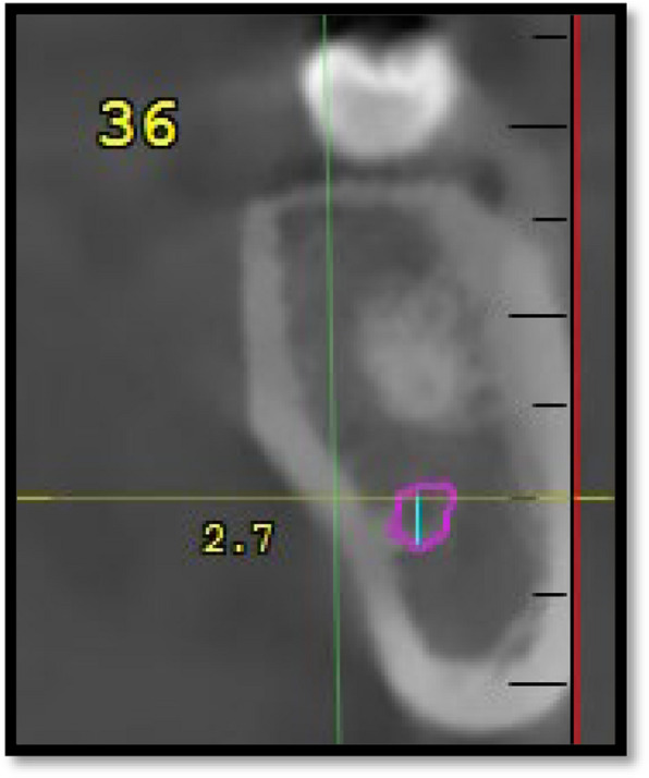
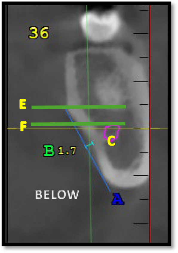
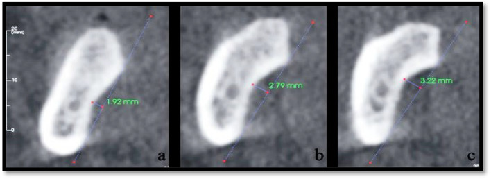
Methodology: CBCT scans of 160 patients (80 males and 80 females) aged 18-35 years were analysed. SF depth was classified into three types: Type I (< 2 mm), Type II (2-3 mm), and Type III (> 3 mm). The MC diameter and concavity angles were measured in the interradicular region of the mandibular molars. The data were statistically analysed via unpaired t tests and chi-square tests (p < 0.05 was considered significant).
Results: Males presented greater mean SF depth, MC diameter, and concavity angles than females did. SF depth was generally more pronounced on the left side in both sexes. Type I SF was the most frequently observed SF depth classification.
Conclusion: CBCT provides valuable insights into mandibular anatomy. Although certain anatomical differences were observed between sexes, particularly in MC diameter, not all findings reached statistical significance. These results suggest the importance of individualized radiographic assessment during surgical planning.

 求助内容:
求助内容: 应助结果提醒方式:


